为了理解右心室(RV)对异常负荷的病理生理学适应机制,实验模型至关重要。然而,评估右心室的尺寸和功能具有复杂性和挑战性。本方案提供了一种对承受右心室压力负荷的小鼠进行心脏磁共振成像(CMR)的方法,作为一种非侵入性的基准检测手段。
方法文章
通过肺动脉束带小鼠模型的心脏磁共振成像对双心室功能与形态的定量分析
Anne-Marie C. Koop*1 , Quint A.J. Hagdorn*1 , Kees (C.) W.A. van de Kolk2,3 , Annemieke van Oosten2 , Michel Weij2 , Herman H.W. Silljé4 , Tineke P. Willems5 , Rolf M.F. Berger1
1Center for Congenital Heart Diseases, Department of Pediatric Cardiology, University Medical Center Groningen, University of Groningen, 2The Central Animal Facility, University Medical Center Groningen, University of Groningen, 3Gronsai (Groningen Small Animal Imaging Facility), University Medical Center Groningen, University of Groningen, 4Department of Cardiology, University Medical Center Groningen, University of Groningen, 5Department of Radiology, University Medical Center Groningen, University of Groningen
* These authors contributed equally
在新标签页中打开
本文内容
摘要
Loading...
$$\rightleftharpoonup{xx}$$
$$\longleftharp{xx}$$,
$$\longrightharp{xx}$$,
摘要
Loading...
$$\rightleftharpoonup{xx}$$
$$\longleftharp{xx}$$,
$$\longrightharp{xx}$$,
右心室(RV)功能与衰竭是获得性和先天性心脏病(包括肺动脉高压)预后的重要决定因素。由于右心室结构复杂,对其功能和形态的评估较为复杂。目前,心脏磁共振(CMR)成像是无创评估右心室功能和形态的金标准。本方案描述了在通过肺动脉缩窄术(PAB)诱导右心室压力负荷的小鼠模型中进行CMR成像的方法。PAB手术通过在23 G针头周围肺动脉上放置6-0缝线完成。使用超声心动图在术后2周和6周测定PAB压差。在术后6周,使用配备1,500 mT/m梯度的9.4 T磁共振成像仪,通过10至11个厚度为1 mm的电影序列切片,测量收缩末期和舒张末期容积及质量,以评估左右心室的形态和功能。代表性结果表明,PAB可显著增加右心室压力负荷,并对双心室形态和右心室功能产生显著影响。同时显示,在持续6周的右心室压力负荷下,心输出量仍得以维持。本文提供了一种可重复的定量评估右心室压力负荷小鼠模型中双心室形态和功能的实验方案,可用于探索右心室重构与功能障碍决定因素的相关研究。
引言
Loading...
$$\rightleftharpoonup{xx}$$
$$\longleftharp{xx}$$,
$$\longrightharp{xx}$$,
患有获得性和先天性心血管疾病的患者,包括肺动脉高压(PH),存在右心室(RV)功能障碍和衰竭的风险1。由于压力负荷增加,右心室适应性改变在早期表现为向心性肥厚,而在疾病晚期则表现为进行性扩张。此外,右心室改变还与代谢异常、细胞外基质紊乱、炎症过程以及最终的右心室衰竭相关2,3,4,5,6。为研究右心室衰竭进展的潜在机制,已建立了多种动物模型。然而,模型的优化以及对右心室功能和结构参数的准确评估仍具挑战性。在无创评估右心室功能和结构方面,心脏磁共振(CMR)成像是金标准。该技术利用强磁场和射频波对跳动的心脏进行成像。CMR已广泛应用于人类,也适用于实验啮齿类动物等动物模型。由于后者心脏体积较小,需要更高的空间分辨率,因此相较于人类,动物成像所需的磁场强度更高。
目前已有多种模拟右心室(RV)压力超负荷的动物模型,包括肺动脉高压(PH)模型7,8,9,10,11,12,13,14,15,16,17以及近端右心室压力负荷模型2,3,10,18,19,20,21,22,23。选择使用PH模型还是近端右心室压力负荷模型,取决于具体的研究问题:若关注干预措施对肺血管系统的影响,从而可能调节右心室后负荷,则应选用PH模型;若关注对右心室的直接作用,则应选用近端右心室压力负荷模型。目前已有多种实验性诱导PH的方法,包括使用野百合碱(MCT)12,13,14,16,22,24,25,26、MCT联合主动脉-下腔静脉分流术9、慢性低氧暴露7,27,28,29,以及血管内皮生长因子受体拮抗剂Sugen 5416联合慢性低氧8,10,30,31。而近端右心室压力负荷模型则属于进行性肺部模型,其并不以肺血管系统为靶点,而是通过缩窄肺动脉造成持续性后负荷增加2,3。该操作可通过缝线结扎(肺动脉结扎,PAB)或在肺动脉周围放置血管夹来实现。PAB已在多种动物物种中实施,并通过多种方法对心脏结构与功能进行了研究,例如组织学、经胸超声心动图(包括斑点追踪技术)以及心导管检查2,32,33,34,35,36,37,38,39,40。在小鼠等小型啮齿类动物中实施PAB具有较大挑战性,因为肺动脉缩窄程度的微小差异即可显著影响右心室压力负荷的程度,进而影响其功能状态和动物存活率。若缩窄过紧,动物可能在术中或术后不久死亡;而若缩窄不足,则无法获得预期的表型。然而,与其它动物相比,小鼠具有显著优势,主要体现在其优异的遗传修饰可行性(如转基因或基因敲除模型)以及繁殖速度快,这对于疾病机制研究以及分子和(表观)遗传因素贡献的探索具有重要价值。
动物研究设计正逐渐转向对疾病过程中时间变化的探究2,3,8,13,21。对于此类研究,无创性检测方法是必要的,因为可以进行系列评估。在评估心脏重构方面,心脏磁共振(CMR)的替代方法可能包括:(1)采用组织病理学进行组织表征,并在不同时间点处死多只动物;(2)通过压力-容积分析进行有创性功能评估;或(3)超声心动图,该方法可使研究者在同一只动物中连续、无创地识别心脏肥厚或扩张。CMR在右心室(RV)评估中具有两大主要优势:(1)CMR是一种无创性检测手段,可在同一只动物中进行多次测量,从而有助于减少实验所需的动物数量;(2)CMR不依赖于特定的几何形状假设,并能实现三维成像。在人类多种心脏疾病中,基于CMR测得的右心室容积和功能参数已被证实具有高准确性,并被视为无创性评估的金标准42,43,44,45,但此前尚未转化为适用于右心室压力超负荷小鼠的CMR检测方案。
文献中描述了多种肺动脉缩窄(PAB)模型,但在评估血流动力学效应以及右心室(RV)功能与适应性的方法上存在高度异质性。本方案概述了小鼠PAB手术的操作流程,并通过超声心动图测量PAB压差,结合心脏磁共振成像(CMR)评估心脏结构和功能,对模型进行验证。尽管已有针对PAB大鼠的CMR检查方案发表,但此类联合方法此前尚未在小鼠中报道。虽然大鼠常用于肺动脉高压(PH)模型8,12,13,14,15,16,22,24,25,26,27,28,29,30,31,46,但小鼠更多用于转基因或基因敲除研究,因而有助于我们深入理解压力超负荷导致右心室衰竭的机制。本方案可为未来探索向右心室衰竭转变过程中相关信号通路的研究奠定基础。
访问受限。请登录或开始试用以查看此内容。
方案
Loading...
$$\rightleftharpoonup{xx}$$
$$\longleftharp{xx}$$,
$$\longrightharp{xx}$$,
所有实验和动物护理均按照荷兰《动物实验法》进行,并符合美国国立卫生研究院发布的《实验动物护理和使用指南》。荷兰格罗宁根大学动物实验委员会已批准本实验方案(许可证编号:2014-041/3005)。
1. 动物饲养与适应性驯化
- 使用20–30 g野生型C57黑6(C57BL/6)小鼠(先前已描述的机构繁育品系47),雌雄不限,所有小鼠均超过8周龄。将小鼠以每笼最多五只的方式群养。为使其适应人为操作,需让小鼠至少适应环境7天。在此期间不得进行任何实验操作。
2. 肺动脉束带手术
- 准备
- 将小鼠放入充满5%异氟烷/100%氧气的诱导室中。通过施加疼痛刺激(例如脚趾夹捏)检查反射是否消失。
- 使用电动剃须刀剃除小鼠左侧半胸壁的毛发。
- 轻轻拉出舌头并施加轻微张力固定。
- 将光源置于声门水平的外部咽喉处,以照亮咽喉内部。
- 使用20 G柔性套管对小鼠进行经气管插管。
- 将动物右侧卧位放置于加热垫上(设定温度为37 °C)。
- 将套管连接至微型呼吸机,开始通气,使用1.5%–2.5%异氟烷/氧气混合气体(呼吸频率180次/分钟,潮气量250 μL)。
- 皮下注射0.1 mg/kg的丁丙诺啡,用于术后镇痛。
- 使用眼膏防止眼睛脱水。
- 经左侧肋间切口行肺动脉束带手术
- 将小鼠右侧卧位固定,右前肢置于中立位,右后肢伸展,左前肢向后弯曲。
- 用氯己定对胸壁皮肤进行消毒,擦拭两次。
- 手术使用无菌器械。用小剪刀(圆柄,12 mm刀片)从左侧腋窝沿第二和第三肋骨方向平行剪开皮肤。
- 辨认胸浅肌(m. pectoralis superficialis,斜行的浅层肌肉)和胸深肌(m. pectoralis profundus,斜行的深层肌肉)。
- 使用缝合环,将胸浅肌向腹侧牵拉,胸深肌向背侧牵拉。
- 打开第二肋间间隙,使用改良的回形针撑开肋骨,使左心耳、左肺和肺动脉清晰可见。
- 分离肺动脉与主动脉。使用钝头25 G针带6-0缝线绕肺动脉放置缝合环,并在肺动脉周围打一个松散的2-1-1结扎。
- 将23 G针平行置于肺动脉旁,并位于6-0缝线内,先固定最靠近近端的缝合结,再固定远端结,随后移除23 G针。确保结扎松紧适当。
- 使用单丝聚丙烯5-0缝线,以两到三个间断缝合关闭胸腔。释放胸浅肌和胸深肌。
- 使用纯聚乙醇酸5-0缝线缝合皮肤。采用连续缝合技术以减少组织瘢痕形成;瘢痕组织会影响超声心动图的图像质量。
- 在恢复麻醉期间继续氧气通气,同时关闭异氟烷,直至小鼠恢复自主呼吸,可通过腹部运动观察到。
- 将气管插管与呼吸机断开。确认出现自主呼吸后方可拔管;若未见自主呼吸,应重新连接呼吸机并返回步骤2.2.12。
- 观察小鼠直至其恢复意识。
- 假手术
- 执行上述操作,但不进行束带结扎(省略步骤2.2.2–2.2.6)。
- 术后阶段
- 术后24小时内将小鼠单独饲养于恒温培养箱中(37 °C)。
- 术后前三天每日观察小鼠状况。如出现任何不适迹象,每日两次皮下注射0.1 mg/kg丁丙诺啡用于术后镇痛。
3. 超声心动图
- 准备
- 在PAB手术后14天,通过超声心动图进行PAB压差分析。
- 启动超声心动图设备,选择心脏检查程序包和14.0 MHz探头。
- 麻醉
- 将小鼠放入充满5%异氟烷和100%氧气混合气体的诱导室中。
- 剃除小鼠胸部的毛发。
- 将小鼠仰卧于加热垫(温度37 °C)上,并将口鼻部置于通气面罩中。
- 以1.5%–2.5%异氟烷与100%氧气(0.15 L/min)及室内空气(0.3 L/min)的混合气体进行通气。
- 通过夹捏脚趾检查麻醉深度,并据此调整麻醉剂量。
- 使用眼膏防止眼睛脱水。
- 通过超声心动图测定PAB压差
- 在每只前肢及两只后肢上分别贴上小儿心电图电极贴片,利用这些贴片固定动物。
- 在小鼠已剃毛的胸部区域涂抹超声耦合剂。
- 获取肺动脉图像时可采用两种切面:胸骨旁长轴(PLAX)或胸骨旁短轴(PSAX)切面。获取两种切面图像,并选择图像质量最佳、流速最高的切面用于分析。
- 获取PLAX和PSAX切面图像。
- 按下彩色多普勒按钮以显示血流情况。
- 将超声探头置于与胸骨旁线成30°角的位置,以获取PLAX切面(详细描述见Cheng等48),显示升主动脉。
- 将探头轻微向左扫查,使升主动脉隐没于肺动脉后方。当可见肺动脉且血流呈垂直方向时,即为正确的PLAX切面。
- 将光标对准肺动脉血流方向。按下连续波(CW)多普勒按钮,记录三个心动周期的流速时间积分测量值。按下保存。
- 从PLAX切面顺时针旋转探头90°以获取PSAX切面,然后将探头轻微向头侧/腹侧倾斜,以获得主动脉水平的PSAX切面。当右心室流出道位于主动脉与探头之间时,即为正确的PSAX切面。该切面中血流继续沿垂直方向进入肺动脉。详细描述见Cheng等48。
- 将光标对准肺动脉血流方向。按下连续波(CW)多普勒按钮,记录三个心动周期的流速时间积分测量值。按下保存。
- 测量最佳切面(PSAX或PLAX)中三个最大流速值并计算其平均值。利用简化的伯努利方程计算PAB压差,单位为毫米汞柱(mmHg)。
4. 心脏磁共振成像
- 准备
- 在PAB手术后6周(即42天)进行CMR分析。
注意:根据研究问题的不同,若需设置多个时间点,可在PAB手术后选择更早的时间点。也可考虑更晚的时间点;然而,右心室衰竭和死亡的发生率可能随之增加。 - 使用足够强的磁铁(通常, >7 T 用于啮齿类动物心脏磁共振扫描。本方案采用9.4 T垂直系统,配备1,500 mT/m梯度组件和89 mm孔径。
- 安装用于分析衍生图像中容积和质量的CMR后处理软件。该软件若支持通过手动分割确定舒张末期(ED)和收缩末期(ES)容积(分别为EDV和ESV)以及心室质量(在ED和ES期均测量),则视为适用。
- 在PAB手术后6周(即42天)进行CMR分析。
- 麻醉与固定
- 将小鼠放入充满5%异氟烷与100%氧气混合气体的诱导室中。通过脚趾夹捏施加疼痛刺激,以确认麻醉效果。
- 在小鼠眼睛上涂抹眼药膏,以保持扫描过程中的眼部湿润。
- 将小鼠放置于扫描仪的动物床中,动物床需集成供气系统,提供含1.5%–2.5%异氟烷、100%氧气(0.15 L/min)和室内空气(0.3 L/min)的预热混合气体(37 °C),并配备压力垫以监测扫描过程中的心率(目标范围:400–500 bpm)和呼吸频率(目标范围:约35次/分钟)。根据这两项参数调节麻醉深度。确保动物床由塑料制成,不含任何磁性材料。
- 将装有小鼠的动物床放入扫描仪中。
- 进行心脏磁共振成像
- 通过调节射频(RF)鸟笼线圈至氢-1(1H)共振频率来进行预采集调整。
- 然后使用自动匀场程序将磁场尽可能调节至均匀。
注意:计算机化匀场通过所谓的调谐方法实现,该方法利用1H自由感应衰减(FID)信号下的面积作为质量参数。在此调谐过程中,系统会以迭代循环方式检查用户定义的一组匀场线圈(Z、Z2、X、Y、XZ和YZ)。依次单独调节每个匀场线圈,以最大化FID信号下的面积。该方法本质上为线性过程,可快速有效地完成匀场。 - 通过调整射频脉冲功率,最大化一维图像轮廓,从而优化射频脉冲。
- 通过使用三平面定位序列(tripilot sequence)进行定位扫描,确定心脏在扫描仪中的精确位置。采用快速梯度回波序列获取胸部的定位图像,包括横断面、冠状面和矢状面切片。图1A,B,C)
- 调整轴线以匹配实际的轴状、两腔和四腔观的轴线图1D,E).
- 随后,将电影序列切面垂直于右心室流出道与右心室最顶端部分之间的假想轴线进行定位。
- 获取10到11个1毫米厚的电影序列切片,切片间无间隙,以完整覆盖右心室从心尖到心底的成像范围。图1F通过自门控IntraGate-快速低角度激发(FLASH)方法实现,该方法无需心电图(ECG)和呼吸门控。采集参数如下所示: 表1将图像保存为 DICOM 格式。
- 对获取的图像进行分析
- 双击软件以打开程序。
- 使用CMR后处理软件打开图像 导入 按钮。
- 确定收缩末期(定义为右心室腔视觉上最小的时相)和舒张末期(定义为右心室腔视觉上最大的时相)。
- 根据心血管磁共振学会的指南49,从心尖到基底部,在舒张末期和收缩末期手动勾画心外膜轮廓,方法是在每幅图像的心外膜边界上标记多个点。在最后一个点处双击以完成心外膜轮廓的绘制。
- 对心内膜轮廓进行相同操作。图2)。左心室和右心室的舒张末期容积(EDV)、收缩末期容积(ESV)、舒张末期质量(ED mass)和收缩末期质量(ES mass)现由软件自动计算。
注意:心肌质量定义为心肌体积乘以心肌密度(即1.05)。 - 根据研究问题和研究人群,使用胫骨长度或体重,依据已发表的公式对受试者体型相关变量进行指数化处理50.
- 在乳头肌中段水平的短轴切面上,分别于舒张末期和收缩末期,将左心室腔平行于室间隔(IVS)的直径除以垂直于室间隔的左心室腔直径,计算偏心率指数(EI)。
- 该软件以 mL 为单位计算每搏输出量(SV)
 ,以及射血分数(EF,%)作为
 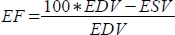 .
. - 以 ml/min 为单位计算心输出量(CO)
 心率通过动物床内置的压力垫手动测量,如上所述,因为扫描仪无法充分记录高频的心率。
 - 根据研究问题和研究人群的不同,可依据已发表的公式,通过胫骨长度或体重对受试者的每搏输出量(SV)和心输出量(CO)进行体表面积校正。50.
5. 统计分析
- 打开用于数据可视化和统计分析的软件。
- 按组(PAB 和 sham)对数据进行排序,每组数据分别置于单独的列中。
- 对每个变量,使用 Mann-Whitney 检验比较 PAB 与 sham 组。
访问受限。请登录或开始试用以查看此内容。
结果
Loading...
$$\rightleftharpoonup{xx}$$
$$\longleftharp{xx}$$,
$$\longrightharp{xx}$$,
PAB 手术的死亡率约为 10%。所展示的结果显示了假手术组(n = 5)和 PAB 组(n = 8)小鼠的特征。如图 3所示,与假手术组动物相比,PAB 组在术后 2 周和 6 周的 PAB 压力梯度值显著升高。这种负荷增加导致右心室扩张,表现为右心室舒张末容积(RV EDV)和收缩末容积(RV ESV)增加(图 4A、B)。右心室功能障碍表现为右心室射血分数(RV EF)下降(图 4C),而右心室每搏输出量(RV SV)保持不变(图 4D)。右心室舒张末质量和收缩末质量均增加,提示出现右心室肥厚(图 4E、F)。左心室舒张末容积(LV EDV)和收缩末容积(LV ESV)减少(图 4G、H
访问受限。请登录或开始试用以查看此内容。
讨论
Loading...
$$\rightleftharpoonup{xx}$$
$$\longleftharp{xx}$$,
$$\longrightharp{xx}$$,
本方案提供了一种在小鼠中进行PAB的可重复方法,以及随后利用CMR评估心脏重构和功能适应的方法。
PAB 与其他增加右心室压力负荷的模型不同,因为它涉及后负荷的绝对且静态的增加,而不伴随其他触发因素。在低氧、野百合碱、分流或这些诱导因素组合的模型中,右心室压力负荷基于肺血管系统的重构。这种重构由内皮损伤、炎症、细胞因子迁移和血管收缩所驱动。这些过程的程度因模型而异,因此压力负荷的程度也随之不同。与这些模型相比,PAB 引起固定的右心室后负荷,因而具有可重复性,并且不受治疗干预的影响。这使得研究人员能够在不影响右心室后负荷的情况下,研究针对压力负荷下右心室的干预措施。小鼠 PAB 模型显示出明显的跨 PAB 压力梯度,从而能够评估这种显著的压力负荷。
由于右心室(RV)呈围绕左心室(LV)的三角形结构,且紧邻胸骨后方,因此通过超声心动图进行容积评估具有挑战性41,42。无论是二维还是三维超声心动图,在准确性方面均...
访问受限。请登录或开始试用以查看此内容。
披露
Loading...
$$\rightleftharpoonup{xx}$$
$$\longleftharp{xx}$$,
$$\longrightharp{xx}$$,
格罗宁根大学医学中心就R.M.F. Berger在本手稿内容之外的咨询活动与Actelion和Lilly公司签订了合同。其他作者声明不存在利益冲突。
致谢
Loading...
$$\rightleftharpoonup{xx}$$
$$\longleftharp{xx}$$,
$$\longrightharp{xx}$$,
我们感谢 P. Da Costa-Martins 在本研究的动物实验中提供的支持。
访问受限。请登录或开始试用以查看此内容。
材料
| 姓名 | 公司 | 目录编号 | 评论 |
|---|---|---|---|
| 14.0 MHz i13L-超声心动图探头 | GE Healthcare, Waukesha, WI, USA | ||
| 20G 套管针 | |||
| 23G 针头 | |||
| 配备 1,500 mT/m 梯度系统的 9.4T 磁共振扫描仪 | Bruker BioSpin, Ellingen, Germany | ||
| 麻醉诱导室 | |||
| 钝头 25G 针头 | |||
| 布托啡诺 | |||
| 氯己定 | |||
| 心脏磁共振后处理软件 | Medis Medical Imaging Systems, Leiden, The Netherlands | Qmass 版本 7.6 | |
| 数据可视化与统计分析软件 | GraphPad Prism Inc, La Jolla, CA, USA | 软件版本 7.02 | |
| 超声心动图设备 | GE Healthcare, Waukesha, WI, USA | Vivid Dimension 7 | |
| 眼药膏 | |||
| 加热垫 | |||
| 孵育箱(37°C) | |||
| 异氟烷 | |||
| 异氟烷蒸发器 | |||
| 啮齿类动物微型呼吸机 | Hugo Sachs | 型号 687 | |
| 单丝聚丙烯 5-0 缝线 | |||
| 单丝聚丙烯 6-0 缝线 | |||
| 皮下注射用针头和注射器 | |||
| 儿科心电图电极贴片 | |||
| 纯聚乙醇酸 5-0 缝线 | |||
| 无菌外科器械 | |||
| 通气面罩 |
参考文献
Loading...
$$\rightleftharpoonup{xx}$$
$$\longleftharp{xx}$$,
$$\longrightharp{xx}$$,
- Norozi, K., et al. Incidence and Risk Distribution of Heart Failure in Adolescents and Adults With Congenital Heart Disease After Cardiac Surgery. The American Journal of Cardiology. 97 (8), 1238-1243 (2006).
- Borgdorff, M. A. J., et al. Clinical symptoms of right ventricular failure in experimental chronic pressure load are associated with progressive diastolic dysfunction. Journal of Molecular and Cellular Cardiology. 79, 244-253 (2015).
- Koop, A. M. C., et al. Right ventricular pressure overload alters cardiac lipid composition. International Journal of Cardiology. , (2019).
- Faber, M. J., et al. Right and left ventricular function after chronic pulmonary artery banding in rats assessed with biventricular pressure-volume loops. American Journal of Physiology and Heart Circirculation Physiology. 291 (4), 1580-1586 (2006).
- Bogaard, H. J., et al. Chronic pulmonary artery pressure elevation is insufficient to explain right heart failure. Circulation. 120 (20), 1951-1960 (2009).
- Samson, N., Paulin, R. Epigenetics, inflammation and metabolism in right heart failure associated with pulmonary hypertension. Pulmonary Circulation. 7 (3), 572-587 (2017).
- Rumsey, W. L., et al. Adaptation to hypoxia alters energy metabolism in rat heart. American Journal of Physiology Heart and Circulatory Physiology. 276 (1), 71-80 (1999).
- Drozd, K., et al. Effects of an endothelin receptor antagonist, Macitentan, on right ventricular substrate utilization and function in a Sugen 5416/hypoxia rat model of severe pulmonary arterial hypertension. Journal of Nuclear Cardiology. 24 (6), 1979-1989 (2017).
- Van Der Feen, D. E., et al. Shunt surgery, right heart catheterization, and vascular morphometry in a rat model for flow-induced pulmonary arterial hypertension. Journal of Visualized Experiments. (120), e55065(2017).
- Gomez-Arroyo, J., et al. Metabolic gene remodeling and mitochondrial dysfunction in failing right ventricular hypertrophy secondary to pulmonary arterial hypertension. Circulation: Heart Failure. 6 (1), 136-144 (2013).
- Bruns, D. R., Dale Brown, R., Stenmark, K. R., Buttrick, P. M., Walker, L. A. Mitochondrial integrity in a neonatal bovine model of right ventricular dysfunction. American Journal of Physiology - Lung Cellular and Molecular Physiology. 308 (2), 158-167 (2015).
- Zhang, W. H., et al. Up-regulation of hexokinase1 in the right ventricle of monocrotaline induced pulmonary hypertension. Respiratory Research. 15 (1), 119(2014).
- Paulin, R., et al. A miR-208-Mef2 axis drives the decompensation of right ventricular function in pulmonary hypertension. Circulation Research. 116 (1), 56-69 (2015).
- Sutendra, G., et al. A metabolic remodeling in right ventricular hypertrophy is associated with decreased angiogenesis and a transition from a compensated to a decompensated state in pulmonary hypertension. Journal of Molecular Medicine. 91 (11), 1315-1327 (2013).
- Balestra, G. M., et al. Increased in vivo mitochondrial oxygenation with right ventricular failure induced by pulmonary arterial hypertension: Mitochondrial inhibition as driver of cardiac failure. Respiratory Research. 16, 6(2015).
- Piao, L., et al. The inhibition of pyruvate dehydrogenase kinase improves impaired cardiac function and electrical remodeling in two models of right ventricular hypertrophy: Resuscitating the hibernating right ventricle. Journal of Molecular Medicine. 88 (1), 47-60 (2010).
- Piao, L., et al. FOXO1-mediated upregulation of pyruvate dehydrogenase kinase-4 (PDK4) decreases glucose oxidation and impairs right ventricular function in pulmonary hypertension: therapeutic benefits of dichloroacetate. Journal of Molecular Medicine. 91, 333-346 (2013).
- Sheikh, A. M., et al. Right ventricular hypertrophy with early dysfunction: A proteomics study in a neonatal model. Journal of Thoracic and Cardiovascular Surgery. 137 (5), 1146-1153 (2009).
- Olivetti, G., et al. Cellular basis of wall remodeling in long-term pressure overload-induced right ventricular hypertrophy in rats. Circulation Research. 63 (3), 648-657 (1988).
- Lauva, I. K., et al. Control of myocardial tissue components and cardiocyte organelles in pressure-overload hypertrophy of the cat right ventricle. The American Journal of Anatomy. 177 (1), 71-80 (1986).
- Fang, Y. H., et al. Therapeutic inhibition of fatty acid oxidation in right ventricular hypertrophy: Exploiting Randle's cycle. Journal of Molecular Medicine. 90 (1), 31-43 (2012).
- Piao, L., et al. Cardiac glutaminolysis: A maladaptive cancer metabolism pathway in the right ventricle in pulmonary hypertension. Journal of Molecular Medicine. 91 (10), 1185-1197 (2013).
- Sack, M. N., Disch, D. L., Rockman, H. A., Kelly, D. P. A role for Sp and nuclear receptor transcription factors in a cardiac hypertrophic growth program. Proceedings of the National Academy of Sciences of the United States of America. 94 (12), 6438-6443 (1997).
- Broderick, T. L., King, T. M. Upregulation of GLUT-4 in right ventricle of rats with monocrotaline- induced pulmonary hypertension. Medical Science Monitor. 14 (12), 261-264 (2008).
- Enache, I., et al. Skeletal muscle mitochondrial dysfunction precedes right ventricular impairment in experimental pulmonary hypertension. Molecular and Cellular Biochemistry. 373 (1-2), 161-170 (2013).
- Sun, X. Q., et al. Reversal of right ventricular remodeling by dichloroacetate is related to inhibition of mitochondria-dependent apoptosis. Hypertension Research. 39 (5), 302-311 (2016).
- Adrogue, J. V., Sharma, S., Ngumbela, K., Essop, M. F., Taegtmeyer, H. Acclimatization to chronic hypobaric hypoxia is associated with a differential transcriptional profile between the right and left ventricle. Molecular and Cellular Biochemistry. 278 (1-2), 71-78 (2005).
- Sharma, S., et al. Dynamic changes of gene expression in hypoxia-induced right ventricular hypertrophy. American Journal of Physiology - Heart and Circulatory Physiology. 286 (3), 1185-1192 (2004).
- Nouette-Gaulain, K., et al. Time course of differential mitochondrial energy metabolism adaptation to chronic hypoxia in right and left ventricles. Cardiovascular Research. 66 (1), 132-140 (2005).
- Graham, B. B., et al. Severe pulmonary hypertension is associated with altered right ventricle metabolic substrate uptake. American Journal of Physiology - Lung Cellular and Molecular Physiology. 309 (5), 435-440 (2015).
- Liu, A., et al. Estrogen maintains mitochondrial content and function in the right ventricle of rats with pulmonary hypertension. Physiological Reports. 5 (6), 1-12 (2017).
- Kobr, J., et al. Right Ventricular Pressure Overload and Pathophysiology of Growing Porcine Biomodel. Pediatric Cardiology. 37 (8), 1498-1506 (2016).
- Yerebakan, C., et al. Acute and chronic response of the right ventricle to surgically induced pressure and volume overload - an analysis of pressure-volume relations. Interactive CardioVascular and Thoracic Surgery. 10 (4), 519-525 (2010).
- Gufler, H., et al. Right Ventricular Function After Pulmonary Artery Banding: Adaptive Processes Assessed by CMR and Conductance Catheter Measurements in Sheep. Journal of Cardiovascular Translational Research. 12 (5), 459-466 (2019).
- Baicu, C. F., et al. Time course of right ventricular pressure-overload induced myocardial fibrosis: relationship to changes in fibroblast postsynthetic procollagen processing. American Journal of Physiology-Heart and Circulatory Physiology. 303 (9), 1128-1134 (2012).
- Manohar, M., et al. Regional myocardial blood flow and coronary vascular reserve in unanesthetized young calves exposed to a simulated altitude of 3500 m for 8-10 weeks. Circulation Research. 50 (5), 714-726 (1982).
- Fávaro, G. A. G., et al. Reversible pulmonary trunk banding: VII. Stress echocardiographic assessment of rapid ventricular hypertrophy in young goats. Journal of Thoracic and Cardiovascular Surgery. 145 (5), 1345-1351 (2013).
- Nielsen, E. A., et al. Regional septal hinge-point injury contributes to adverse biventricular interactions in pulmonary hypertension. Physiological Reports. 5 (14), 1-13 (2017).
- Borgdorff, M. A., et al. Sildenafil enhances systolic adaptation, but does not prevent diastolic dysfunction, in the pressure-loaded right ventricle. European Journal of Heart Failure. 14 (9), 1067-1074 (2012).
- Gold, H., Prindle, K., Levey, G., Epstein, S. Effects of experimental heart failure on the capacity of glucagon to augment myocardial contractility and activate adenyl cyclase. The Journal of Clinical Investigation. 49 (5), 999-1006 (1970).
- Brittain, E. L., et al. Right ventricular plasticity and functional imaging. Pulmonary Circulation. 2 (3), 309-326 (2012).
- Jiang, L., et al. Three-dimensional Echocardiography In Vivo Validation for Right Ventricular Volume and Function. Circulation. 89, 2342-2350 (1994).
- Markiewicz, W., Sechtem, U., Higgins, C. B. Evaluation of the right ventricle by magnetic resonance imaging. American Heart Journal. 113 (1), 8-15 (1987).
- Pattynama, P. M. T., et al. Reproducibility of MRI-derived measurements of right ventricular volumes and myocardial mass. Magnetic Resonance Imaging. 13 (1), 53-63 (1995).
- Wiesmann, F., et al. Comparison of fast spiral, echo planar, and fast low-angle shot MRI for cardiac volumetry at .5T. Journal of Magnetic Resonance Imaging. 8 (5), 1033-1039 (1998).
- Van der Feen, D. E., et al. Multicenter Preclinical Validation of BET Inhibition for the Treatment of Pulmonary Arterial Hypertension. American Journal of Respiratory and Critical Care Medicine. 200 (7), 910-920 (2019).
- da Costa Martins, P. A., et al. MicroRNA-199b targets the nuclear kinase Dyrk1a in an auto-amplification loop promoting calcineurin/NFAT signalling. Nature Cell Biology. 12 (12), 1220-1227 (2010).
- Cheng, H. W., et al. Assessment of right ventricular structure and function in mouse model of pulmonary artery constriction by transthoracic echocardiography. Journal of Visualized Experiments. (84), e51041(2014).
- Schulz-Menger, J., et al. Standardized image interpretation and post processing in cardiovascular magnetic resonance: Society for Cardiovascular Magnetic Resonance (SCMR) Board of Trustees Task Force on Standardized Post Processing. Journal of Cardiovascular Magnetic Resonance. 15 (1), 1-19 (2013).
- Hagdorn, Q. A. J., et al. A novel method optimizing the normalization of cardiac parameters in small animal models: The importance of dimensional indexing. American Journal of Physiology - Heart and Circulatory Physiology. 316 (6), 1552-1557 (2019).
- Scherrer-Crosbie, M., et al. Determination of Right Ventricular Structure and Function in Normoxic and Hypoxic Mice. Circulation. 98 (10), 1015-1021 (2012).
- Wiesmann, F., et al. Analysis of right ventricular function in healthy mice and a murine model of heart failure by in vivo MRI. American Journal of Physiology-Heart and Circulatory Physiology. 283 (3), 1065-1071 (2002).
- Lu, X., et al. Accuracy and Reproducibility of Real-Time Three-Dimensional Echocardiography for Assessment of Right Ventricular Volumes and Ejection Fraction in Children. Journal of the American Society of Echocardiography. 21 (1), 84-89 (2008).
- Soriano, B. D., et al. Matrix-array 3-dimensional echocardiographic assessment of volumes, mass, and ejection fraction in young pediatric patients with a functional single ventricle: A comparison study with cardiac magnetic resonance. Circulation. 117 (14), 1842-1848 (2008).
- Damy, T., et al. Prevalence of, associations with, and prognostic value of tricuspid annular plane systolic excursion (TAPSE) among out-patients referred for the evaluation of heart failure. Journal of Cardiac Failure. 18 (3), 216-225 (2012).
- Kowalik, E., Kowalski, M., Rózański, J., Kuśmierczyk, M., Hoffman, P. The impact of pulmonary regurgitation on right ventricular regional myocardial function: An echocardiographic study in adults after total repair of tetralogy of fallot. Journal of the American Society of Echocardiography. 24 (11), 1199-1204 (2011).
- Koestenberger, M., et al. Systolic right ventricular function in pediatric and adolescent patients with tetralogy of Fallot: Echocardiography versus magnetic resonance imaging. Journal of the American Society of Echocardiography. 24 (1), 45-52 (2011).
- Bovens, S. M., et al. Evaluation of infarcted murine heart function: Comparison of prospectively triggered with self-gated MRI. NMR in Biomedicine. 24 (3), 307-315 (2011).
访问受限。请登录或开始试用以查看此内容。
重印与许可
申请许可以重复使用本 JoVE 文章的文本或图表
申请许可标签