Guest Editors
Selwin Wu
National University of Singapore, Department of Biological Sciences, Mechanobiology Institute
<p>Selwin graduated with First-Class Honours and a PhD with Dean's Award from the University of Queensland, Australia. During his post-doctoral training at Harvard Medical School, HHMI, and Dana-Farber Cancer Institute, he received a Leukemia & Lymphoma Society Fellow Award. He then returned to Singapore and was awarded a Young Individual Research Grant by the National Medical Research Council.</p> <p>He investigates how processes observed in tumors and development, including cell invasion and extrusion, emerge from changes in gene regulation, cellular interactions, signalling, mechanics, and architecture. His research applies gene expression analysis, micropatterning, molecular biology techniques, and a variety of photonic methods to study two and three-dimensional cell cultures.</p>
Nicholas Ariotti
The University of New South Wales, School of Medical Sciences, Mark Wainwright Analytical Centre
<p>Dr. Nicholas Ariotti began his research career at the University of Queensland where he completed a PhD in Cell and Membrane Biology at the Institute for Molecular Biosciences. During this time, he used diverse electron microscopy-based techniques to understand the structure, function, and composition of specialized and endocytic plasma membrane microdomains termed caveolae. Since then, Dr. Ariotti’s research has focused on developing novel correlative light and electron microscopy methods to understand how proteins and lipids are organized at the cell surface and in cellular trafficking. In 2017 he took up an Associate Director position at the Electron Microscope Unit at The University of New South Wales. He has published many peer-reviewed articles in journals including <em>Nature, Cell, Nature Cell Biology, Nature Nanotechnology, Nature Communications, Current Biology, The Journal of Cell Biology, </em>and<em> Developmental Cell</em>.</p>
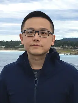
Shiwei Liu
Harvard University, USA
<p>Shiwei Liu received his PhD in Biological and Biomedical Sciences from Harvard University, Harvard Medical School. During his thesis work, he investigated the mechanisms controlling nuclear envelope formation and how defects in nuclear envelope formation cause genome instability and nuclear dysfunction. Currently, he is working as a postdoc researcher at Harvard University. He is studying the 3D genome organization during cell fate specification in Professor Zhuang Xiaowei’s laboratory.</p>
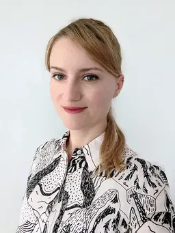
Joyce Meiring
Utrecht University, Netherlands
<p>Joyce Meiring obtained a bachelor in biomedical science with first class honors from University of Queensland, by then the cytoskeleton had already peaked her interests. She completed her PhD in 2018 at University of New South Wales working on the organization and recruitment of the tropomyosin family of actin-regulators where she received a Dean’s Award for Outstanding PhD Theses. She is currently doing a postdoc under an EMBO fellowship at the University of Utrecht, probing the biomechanical roles of microtubules using light-switchable tools.</p>
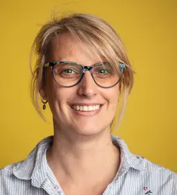
Anne Lagendijk
University of Queensland, Institute for Molecular Bioscience, Australia
<p>Dr. Anne Lagendijk’s laboratory at the Institute for Molecular Biosciences, University of Queensland uses zebrafish as a genetic model that is extremely suitable for live imaging, with a platform for generating 3D bioengineered human micro-vessels.</p> <p>These models allow them to uncover the mechanisms that are essential for endothelial cell function in lumenized vessels that are under flow pressure and are exposed to physiologically relevant extracellular cues. In zebrafish they apply technically challenging genetic, transgenic, and live-imaging approaches that probe endothelial cell function live, up to level of single cells.</p> <p>In addition, their recently implemented platform of bioengineered human vasculature complements the zebrafish work and allows them to tune and test parameters that contribute to endothelial cell biology, like blood flow and ECM stiffness.</p>
Collection Overview
The physical and chemical properties of living cells and tissues dictate biological form and function. Dysregulation of these properties leads to disease such as cancer, neurological disorders, and cardiovascular disease. A clearer understanding of both biophysical and biochemical properties requires methodologies ranging from computational analysis to a variety of photonic methods. For example, combining high-throughput microscopy of biochemical biosensors and automated image analysis would accelerate our quantitation of big data generated from complex multi-layered tissues during development. Thus, improving our understanding of how biochemical signals are transduced into forces that shape organs. In this collection, we seek out articles that would provide clear and detailed demonstration of biochemical, biophysical, and computational approaches that have strong potential to uncover new biological insights. We believe that audio-visual demonstration of protocols would help make these methods more widely accessible and robustly reproducible.
We welcome electron/light microscopists, biophysicists, biochemists, and computational scientists to contribute their methods paper to this issue in the broad area of molecular, cell, developmental biology and related diseases.
Articles