We describe methods for isolating mouse ovarian somatic cells from a stroma-enriched fraction of the ovary and for generating mouse ovarian somatic organoids using a scaffold-free approach.
Method Article
Probing the Ovarian Microenvironment: Methods for Isolation, 2D Culture, and Organoid Culture of Mouse Ovarian Somatic Cells
Opens in a new tab
In This Article
Summary
Loading...
$$\rightleftharpoonup{xx}$$
$$\longleftharp{xx}$$,
$$\longrightharp{xx}$$,
Abstract
Loading...
$$\rightleftharpoonup{xx}$$
$$\longleftharp{xx}$$,
$$\longrightharp{xx}$$,
The ovary consists of heterogeneous populations of somatic cells, both within the follicle and the surrounding stroma, which are critical to support ovarian function and for the generation of high-quality gametes. We report methods for isolating somatic cells from mouse ovaries, including endothelial, epithelial, steroidogenic, stromal, and immune cells. When these primary ovarian somatic cells are plated and cultured in a traditional 2D culture system, the cellular heterogeneity, organization, as well as cell-cell and cell-matrix interactions typically found in the ovary are lost. Thus, we also describe how to generate mouse ovarian somatic organoids using a scaffold-free approach. These organoids self-assemble, maintain diverse cell populations, and produce extracellular matrix and secreted factors, including cytokines. Organoids can be utilized for co-culture experiments and can be maintained in culture for at least 3 weeks with high viability. Overall, these models enable interrogation of ovarian physiology and pathology from the somatic cell perspective.
Introduction
Loading...
$$\rightleftharpoonup{xx}$$
$$\longleftharp{xx}$$,
$$\longrightharp{xx}$$,
Although the oocyte, or female gamete, is essential for giving rise to the next generation, somatic cell populations in the ovary play critical roles in gamete development, steroid hormone production, and ovarian homeostasis. Oocytes, surrounded by somatic granulosa and theca cells, form the functional units of the ovary known as follicles. Granulosa cells are physically connected to the oocyte by transzonal projections and provide metabolic and nutrient support to the oocyte in addition to regulating meiotic arrest1,2. Moreover, in response to gonadotropins, granulosa and theca cells produce steroid hormones, including estrogen, progesterone, and androgens, which have effects beyond the reproductive system, impacting the cardiovascular, musculoskeletal, and nervous systems, and thereby affecting overall female health3. Ovarian follicles develop within a heterogeneous ovarian stroma composed of vasculature, immune cells, several populations of interstitial fibroblasts and mesenchymal cells, as well as a highly ordered extracellular matrix (ECM)4. This stromal microenvironment provides physical and chemical cues that impact follicle growth, ovulation, and luteinization4.
Several methods exist for isolating and culturing gametes and follicles in vitro, and these methods have advanced our understanding of oogenesis and folliculogenesis5. However, the somatic compartment of the ovary is relatively understudied, in part, due to the lack of robust in vitro models. Current models of the ovarian somatic compartment are largely restricted to a single cell type, such as fibroblasts, ovarian surface epithelial, endothelial, immune, or granulosa cells6,7,8,9,10,11. The culture of broadly defined primary ovarian somatic cells in 2D favors macrophage populations over time, resulting in a loss of cellular heterogeneity12. Moreover, traditional two-dimensional (2D) monolayer culture does not allow for the diverse cell-cell and cell-matrix interactions that are present in the ovary. Ovarian tissue explants maintain the cellular and structural complexity of the native tissue but vary in composition depending on where within the ovary they were derived13,14.
Organoids are three-dimensional (3D), multi-cellular, miniaturized versions of organs or tissues that recapitulate in vivo organization and functions15,16. Organoids can be generated from stem cells or primary cells isolated from normal or diseased tissue15. In the context of the female reproductive system, organoids have been generated to model the endometrium, fallopian tube, cervix, and placenta17. However, with respect to the ovary, until recently, organoid models have been limited to a single cell type, such as ovarian surface epithelial or cancer cells, or have been generated from induced pluripotent stem cells to resemble follicle structures rather than the ovarian stroma17,18. Recent studies have generated and characterized organoids using heterogeneous populations of somatic cells isolated from mouse, rhesus macaque, and human ovarian tissue19,20,21. These organoid models have enabled investigation of cellular mechanisms underlying ovarian aging, as well as the effects of phthalate exposure on ECM composition19,21.
Given the critical importance of somatic cells, both in the follicle and stroma, to ovarian function, we describe methods for isolating somatic cells from a stroma-enriched fraction of mouse ovaries and for culturing them in vitro using either traditional monolayer culture or in an organoid model. Using scaffold-free agarose micromolds, ovarian somatic cells form organoids within 1–3 days. Organoids preserve key ovarian cell populations, including fibroblasts, macrophages, and steroidogenic cells, and can be cultured for up to 3 weeks with high viability. Ovarian somatic organoids are conducive to transcriptomic and histologic assessments, analysis of conditioned media, co-culture with other cell types, and compound screening methods. Overall, these organoids are a robust model for in vitro interrogation and modulation of the ovarian somatic compartment and can be applied to the study of ovarian development, aging, physiology, and pathology.
Access restricted. Please log in or start a trial to view this content.
Protocol
Loading...
$$\rightleftharpoonup{xx}$$
$$\longleftharp{xx}$$,
$$\longrightharp{xx}$$,
The use of animals to develop this protocol was in accordance with the Institutional Animal Care and Use Committee at Northwestern University and the National Institutes of Health Guidelines for Care and Use of Laboratory Animals (Northwestern University Animal Welfare Assurance Number: A3283-01; IACUC Protocol ID: IS00013082_IM12). Manufacturer information and catalog numbers for all materials, including silicone casts for agarose micromolds, media, and supplements, are given in the Table of Materials. An overview of the protocol steps is presented in Figure 1. All protocol steps should be performed using appropriate personal protective equipment, in compliance with all institutional biosafety and environmental health and safety regulations, and, unless otherwise specified, within a Class II biosafety cabinet.
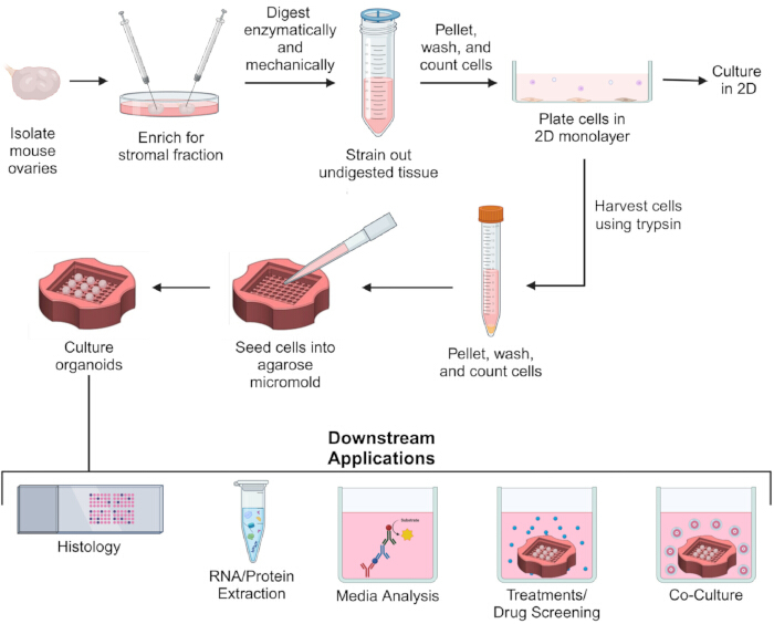
Figure 1: Schematic of mouse ovarian somatic cell isolation, ovarian somatic organoid generation, and downstream applications. Mouse ovaries are isolated and the stromal fraction is enriched. The enriched stromal fraction is digested enzymatically and mechanically. Undigested tissue is strained out. Cells are pelleted, washed, counted, and plated for culture in 2D. For organoid generation, following 2D culture overnight, cells are harvested using trypsin and are pelleted, washed, and counted. Cells are seeded into agarose micromolds for organoid culture. Organoids can be utilized for histology, RNA/protein extraction, conditioned media analysis, treatments, and drug screening, as well as co-culture. Please click here to view a larger version of this figure.
1. Preparation of media for ovarian somatic cell isolation and culture
NOTE: Digestion of ovarian tissue and subsequent isolation and culture of primary somatic cells requires four types of media: Dissection media (DM; used in Steps 2.3–2.4 for dissection and puncture of mouse ovaries), enzymatic media (EM; used in Steps 2.5–2.8 for enzymatic digestion of stroma-enriched mouse ovaries), quenching media (QM; used in Step 2.9 to slow enzymatic digestion), and plating media (PM; used in Steps 2.11–2.14 and Step 4.6 for washing, plating, and 2D culture of primary ovarian somatic cells, as well as inactivation of trypsin when harvesting the cells from 2D culture). All media should be prepared in a sterilized, Class II biological safety cabinet or laminar flow hood. Media recipes listed below are sufficient for processing ovaries from 5–6 mice, but should be scaled as needed. All media can be prepared up to a week in advance and stored at 4 °C, but the enzymes for the EM must be added fresh on the day of cell isolation.
- Prepare DM (15 mL) containing 1% fetal bovine serum (FBS) and 0.5% penicillin-streptomycin (PS) in Leibovitz's L-15 medium. Sterilize the media using a sterile filter.
NOTE: DM is buffered for ambient air exchange and utilized during microdissection of the ovary from surrounding tissues, as well as enrichment for the stromal fraction. - Prepare EM (10 mL) containing 1% FBS and 0.5% PS in αMEM. Add 82 enzymatic units/mL Collagenase IV and 0.2 mg/mL of DNase I (≥400 enzymatic activity units/mg). Dissolve the enzymes by inverting and then sterilize the media using a sterile filter.
NOTE: Enzymatic media is sodium bicarbonate-buffered for air exchange at 5% CO2 and utilized for enzymatic digestion of ovarian tissue pieces. - Prepare QM (20 mL) containing 10% FBS and 0.5% PS in αMEM medium. Sterilize the media using a sterile filter.
NOTE: QM is sodium bicarbonate-buffered for air exchange at 5% CO2 and utilized for quenching of the enzymatic digestion. - Prepare PM (30 mL) containing 10% FBS and 1% PS in RPMI 1640 (containing 25 mM HEPES and 2 mM L-Glutamine). Sterilize the media using a sterile filter.
NOTE: PM is sodium bicarbonate-buffered for air exchange at 5% CO2 and utilized for plating of primary ovarian somatic cells, as well as culture in 2D.
2. Digestion of ovarian tissue and isolation of primary somatic cells
NOTE: Mouse ovaries are isolated and digested both enzymatically and mechanically to isolate primary somatic cells. Ovaries from 5–6 mice can be processed together in the same dish. The following steps should be performed using aseptic technique, preferably using a dissecting microscope in a laminar flow hood.
- Add 10 mL of EM, 20 mL of QM, and 20 mL of PM into separate 100 mm Petri dishes. Equilibrate the dishes in a tissue culture incubator (humidified environment, 37 °C with 5% CO2) for a minimum of 1 h prior to use.
- Warm DM in a water bath (37 °C) prior to use.
- Dissect mouse ovaries into a 35 mm Petri dish containing 3 mL of DM. Separate the ovaries from the attached adipose tissue, oviduct, and ovarian bursa using fine-tipped forceps and/or microscissors (Figure 2Ai,ii). Utilize a dissecting microscope as needed for microdissection.
- Transfer the dissected ovaries to a 45 mm watch glass with 2–3 mL of DM. Under a dissecting microscope, anchor and puncture all areas of the ovaries using 28-G insulin needles with the nondominant and dominant hands, respectively, for approximately 10 min. Frequently rotate the ovarian tissue to ensure that all areas are disrupted.
NOTE: Puncturing the ovaries enriches the stromal fraction by releasing preantral follicles as well as cumulus-oocyte-complexes and granulosa cells from antral follicles (Figure 2Aiii–iv). - Use fine-tipped forceps to move stroma-enriched mouse ovaries to the Petri dish with equilibrated EM (Figure 2Bi).
- Working quickly to minimize temperature and pH changes to the media, tear the stroma-enriched ovaries into approximately 2–3 mm pieces with fine-tipped forceps (Figure 2Bii).
- Hold the Petri dish at an angle and repeatedly pipette EM containing ovarian tissue pieces with a p1000 pipette, set to 1 mL, 100× or for 3 min, whichever comes first (Figure 2Biii). Swirl the Petri dish as necessary to make sure that all tissue pieces are pipetted.
NOTE: If the tissue is too large to be pipetted with a p1000 tip, the tip can be cut with a sterile scalpel to make the opening wider. The bore should be wide enough to allow most (>50%), but not all, of the tissue pieces to enter, creating a shearing effect. - Transfer the Petri dish containing EM and ovarian tissue pieces to a tissue culture incubator (humidified environment, 37 °C with 5% CO2) and incubate for a total of 30 min, removing the dish and pipetting repeatedly (100× or for 3 min) after 15 min, and then again at the end of the 30 min incubation (Figure 2Biii).
NOTE: After the first 15 min incubation, the tissue pieces should be digested enough so that >80% fit through a p1000 pipette tip without the need to cut the tip to make the opening wider. The total duration for the digestion and the amount of pipetting required will be dependent on the starting size of the torn tissue pieces. Furthermore, the age and strain of the mouse, lot-to-lot variability in the activity of the enzymes, and the pipette bore size can all impact the rate of tissue breakdown. Digestion should be quenched when the ovarian tissue pieces are broken down into approximately 1 mm fragments that are still visible by eye (Figure 2Biv). Over-digestion may result in cell loss and decreased cell viability. - Dilute the enzymes to slow tissue digestion by adding 20 mL of preequilibrated QM (Figure 2Ci).
- Pass the cell suspension through a 40 µm cell strainer placed over a 50 mL conical tube to remove undigested tissue pieces (Figure 2Cii). Centrifuge the filtrate at 300 × g for 5 min at room temperature to pellet the cells (Figure 2Ciii).
- Wash the cells by removing the supernatant by aspiration, resuspending the cell pellet in 1 mL of preequilibrated PM, transferring the cell suspension to a 1.5 mL microcentrifuge tube, and centrifuging at 300 × g for 5 min at room temperature to pellet the cells again (Figure 2Civ).
- Remove the supernatant, resuspend the cell pellet in 1 mL of PM, stain a 10 µL aliquot of cells with trypan blue to assess cell viability, and count the cells using a hemocytometer.
NOTE: See Table 1 for anticipated numbers of primary ovarian somatic cells, depending on mouse strain and age, at this stage of the isolation protocol. Cell viability at this stage should be >80%. - Plate the cells on a tissue culture-treated vessel at approximately a density of 1.25 × 10⁵-1.5 × 10⁵/ cm2 (e.g., for cell counts of approximately 3 × 10⁶, plate cells onto a tissue culture-treated 60 mm dish, and for cell counts of approximately 1 × 10⁶, plate cells onto one well of a tissue culture-treated 6-well plate).
- Add additional PM depending on the total volume of the plate or dish (e.g., add an additional 4 mL of PM if using a 60 mm dish or an additional 2 mL of PM if using a well of a 6-well plate).
NOTE: The plating density listed above is recommended if the cells will be utilized for organoid generation following 2D culture overnight. For a longer 2D culture, cells can be plated at a lower density. Extra plating media that has not been equilibrated can be stored at 4 °C for up to 1 week. PM is required both for the generation of organoids and to maintain ovarian somatic cells in 2D culture. At this point in the protocol, the ovarian somatic cells can be maintained in a 2D monolayer culture (step 2.14) or can be used to generate organoids (proceed to Section 3). 2D culture overnight prior to organoid generation is necessary to remove any oocytes from early-stage follicles that may have passed through the 40 µm cell strainer.
- Add additional PM depending on the total volume of the plate or dish (e.g., add an additional 4 mL of PM if using a 60 mm dish or an additional 2 mL of PM if using a well of a 6-well plate).
- Following plating overnight (minimum 16 h), wash the cells with phosphate-buffered saline (PBS; Mg/Ca-free, pH 7.0–7.3) to remove dead cells and debris (Figure 3A). Add fresh PM following washing and replace media with fresh PM every other day throughout the culture (Figure 3B).
NOTE: Despite high cell viability following isolation, a large attrition in total cell count is typically observed after plating overnight due either to cell death or inability to attach to the tissue culture vessel (Figure 3A).
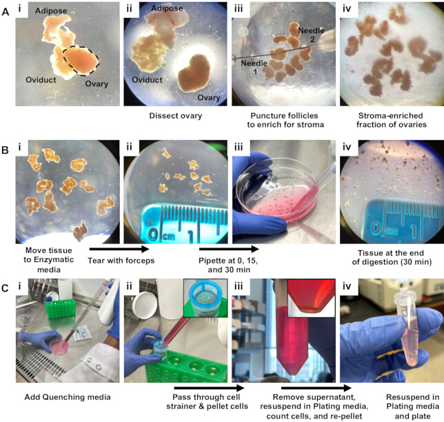
Figure 2: Schematic of mouse ovarian somatic cell isolation. (A) Representative images of (i,ii) mouse ovary dissection and (iii,iv) stroma-enrichment. The mouse ovary is outlined with a dashed line in Ai. (B) Representative images of ovarian tissue pieces throughout mechanical and enzymatic digestion. Stroma-enriched ovaries (i) before and (ii) after tearing, (iii) ovarian tissue pieces during pipetting, and (iv) ovarian tissue pieces after digestion. (C) Representative images of (i) quenching the digestion, (ii) filtering the cell suspension, and (iii,iv) pelleting the primary mouse ovarian somatic cells. Insets in Cii and Ciii show undigested tissue pieces caught by the cell strainer and cell pellet following centrifugation, respectively. Please click here to view a larger version of this figure.
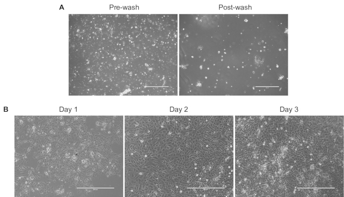
Figure 3: Primary ovarian somatic cells can be cultured in 2D. (A) Representative transmitted light images of primary ovarian somatic cells plated in a 60 mm dish 16 h after plating, before and after washing with PBS. (B) Representative transmitted light images of primary ovarian somatic cells plated in a 24-well plate over 3 days in culture. Scale bars = 400 µm. Please click here to view a larger version of this figure.
3. Preparation of agarose micromolds and media for organoid generation and culture
NOTE: Ovarian somatic organoids are generated and cultured in scaffold-free agarose micromolds with ovarian organoid media. Agarose micromolds and ovarian organoid media should be prepared using aseptic technique in a sterilized, Class II biological safety cabinet or laminar flow hood.
- Preparation of agarose micromolds
- Place silicone casts for micromolds and Graefe forceps into a sterilization pouch and autoclave using a dry cycle to sterilize (Jacket pressure: 20 psi, Chamber temperature: 250 °F (121.11 °C, Sterilizing time: 15 min).
NOTE: Several silicone casts can be autoclaved and used at one time for more rapid generation of agarose micromolds. - Add 1 mL of PBS containing 1% PS to each well of a 24-well tissue culture plate for storage of the agarose micromolds.
- Dissolve 1.5% (w/v) sterile agarose in PBS by heating in a microwave, in a hot water bath, or on a hot plate.
NOTE: Dissolving the agarose by microwaving in a 50 mL conical tube placed in a 100 mL beaker filled with water works well (Figure 4Ai). In addition to allowing for controlled boiling, the hot water bath created in the beaker slows the re-solidification of the agarose. - Allow the liquid agarose to cool to approximately 50 °C until the tube can be comfortably handled.
- Slowly pipette 300–350 µL of 1.5% agarose into the silicone cast, while making sure not to overfill or underfill the cast and avoiding creating bubbles (Figure 4Aii). Make sure the agarose creates a flat layer at the top of the cast rather than a concave or convex edge (Figure 4Aiii,iv). Pop bubbles using a sterile p10 pipette tip before the agarose starts to solidify (Figure 4Bi–iii).
- Allow the agarose to solidify in the silicone cast for 2–3 min at room temperature (Figure 4Biv).
- Once solid, invert the silicone cast over a well of the 24-well tissue-culture plate containing PBS + 1% PS. Carefully flex the silicone to release the agarose micromold into the plate (Figure 4Ci).
NOTE: Agarose micromolds submerged in PBS +1% PS in a 24-well tissue culture plate wrapped with parafilm can be stored at 4 °C up to 1 month prior to use (Figure 4Cii). Agarose micromolds should be prepared at least 16 h prior to use, as microwells often contain air bubbles immediately following casting and submergence in PBS + 1% PS (Figure 4Ciii). However, these air bubbles dissipate during storage (Figure 4Civ). Prior to use for organoid culture, agarose microwell inserts should be carefully examined under a microscope to confirm that microwells were not merged or disrupted during casting (Figure 4Di–iv). Defective micromolds are not recommended for the generation and culture of organoids. It is recommended that an excess of agarose micromolds be prepared to account for ~25% that will have defects
- Place silicone casts for micromolds and Graefe forceps into a sterilization pouch and autoclave using a dry cycle to sterilize (Jacket pressure: 20 psi, Chamber temperature: 250 °F (121.11 °C, Sterilizing time: 15 min).
- Preparation of ovarian organoid media
- In a biosafety cabinet, add organoid growth medium (Mouse) supplements 1 and 2 and 1% PS to basal medium per the manufacturer's instructions. Store the prepared ovarian organoid media at 4 °C for up to 2 weeks prior to use.
NOTE: Supplements 1 and 2 can be aliquoted and stored at -20 °C.
- In a biosafety cabinet, add organoid growth medium (Mouse) supplements 1 and 2 and 1% PS to basal medium per the manufacturer's instructions. Store the prepared ovarian organoid media at 4 °C for up to 2 weeks prior to use.
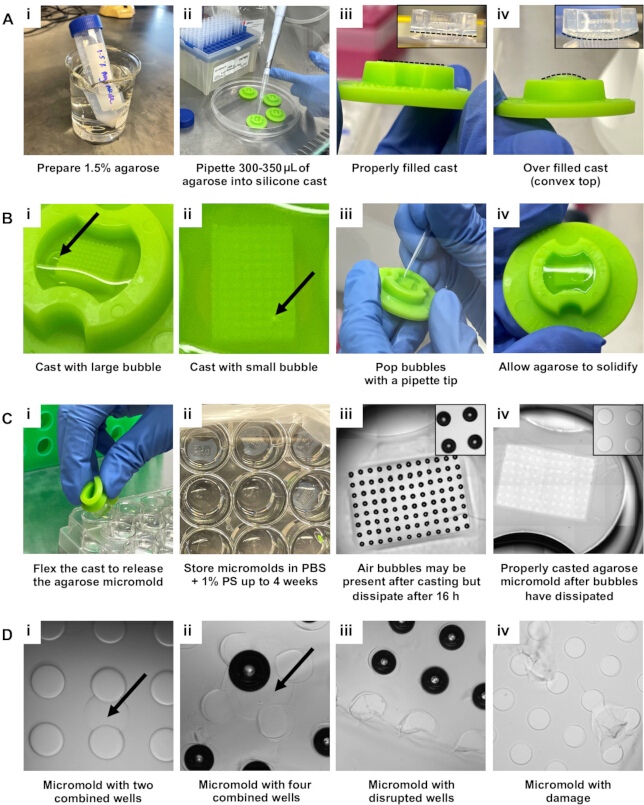
Figure 4: Schematic displaying preparation of agarose micromolds. (A) Representative images of (i) 1.5% agarose preparation, (ii) pipetting into silicone casts, (iii) properly filled, and (iv) overfilled silicone casts. Dashed lines in Aiii and Aiv show flat and convex bottoms of agarose micromolds, respectively. (B) Representative images of (i,ii) filled silicone casts with bubbles in the agarose, (iii) popping bubbles with a pipette tip prior to agarose solidification, and (iv) a solidified agarose micromold within the silicone cast. (C) Representative images of (i) removal of solidified agarose micromolds from silicone casts, (ii) storage of agarose micromolds in 24-well plate containing PBS + 1% PS, (iii) bubbles present in microwells immediately after casting, and (iv) bubble-free, properly casted agarose micromold after 16 h of storage. (D) Representative images of micromolds with (i,ii) combined wells, (iii) disrupted wells, or (iv) damage. Abbreviations: PBS = phosphate-buffered saline; PS = penicillin-streptomycin. Please click here to view a larger version of this figure.
4. Generation and culture of organoids
NOTE: Following 2D culture overnight, primary ovarian somatic cells are harvested from the 2D culture and seeded into agarose micromolds to generate organoids. The following steps should be performed using aseptic technique in a sterilized, Class II biological safety cabinet.
- Warm PM and organoid media to 37 °C in a water bath and warm 0.05% Trypsin to room temperature.
- Using Graefe forceps, carefully transfer the micromolds into the wells of a new 24-well plate (Figure 5Ai).
- Add 500 µL of ovarian organoid media to each well of the 24-well plate containing an agarose micromold (Figure 5Aii). Make sure that micromolds are fully submerged in media and equilibrate the micromolds and media for at least 30 min in an incubator (humidified environment, 37 °C with 5% CO2).
- Remove PM from adherent ovarian somatic cells by aspirating and wash the cells with Mg/Ca-free PBS.
- Add 0.05% Trypsin (1–2 mL of trypsin for a 6-well plate or 60 mm dish) to harvest the cells from 2D culture. Incubate the cells with trypsin for 2–3 min at 37 °C in an incubator.
- Confirm that cells have detached under a microscope and inactivate the trypsin by adding two volumes of warmed PM for every one volume of trypsin used.
- Transfer the cell suspension to a 15 mL conical tube, and pellet the cells by centrifugation at 300 × g for 5 min at room temperature.
- Resuspend the cells in 1 mL of warmed PM, stain an aliquot of cells with trypan blue to assess cell viability, and count the cells using a hemocytometer.
NOTE: See Table 1 for anticipated numbers of primary ovarian somatic cells, depending on mouse strain and age, following 2D culture overnight and subsequent trypsinization. Viability of trypsinized cells is typically greater than 90%. - Pellet the cells by centrifugation (300 × g for 5 min at room temperature) and resuspend the cell pellet in ovarian organoid media to a density of 3.33 × 10⁶ live cells per 1 mL of media, i.e., 2.5 × 10⁵ live cells per 75 µL of media.
NOTE: Here, 2.5 × 10⁵ cells are seeded per agarose micromold. Thus, the total number of micromolds that can be generated will depend on the total cell count. Organoids can be generated with fewer than 2.5 × 10⁵ cells per agarose micromold, but organoid size, aggregation kinetics, relative cellular composition, and functions, including hormone production, may be impacted. - Remove the equilibrated agarose micromolds from the incubator, make sure that the micromolds are oriented such that the cell seeding chamber is facing up, and remove the ovarian organoid media from the cell seeding chambers (Figure 5Aiii).
- Remove the media from the cell seeding chambers of the micromolds by using Graefe forceps to lift and tilt the micromolds or by pipetting. To remove media from cell seeding chambers by pipetting, gently hover the pipette tip over the corner of the cell seeding chamber while drawing up media. Do not make direct contact with the agarose micromold to avoid disrupting the microwells.
NOTE: Cell seeding chambers should be as dry as possible, such that microwells are visible by eye, but the outside of the micromolds should still be surrounded by ovarian organoid media (Figure 5Aiii).
- Remove the media from the cell seeding chambers of the micromolds by using Graefe forceps to lift and tilt the micromolds or by pipetting. To remove media from cell seeding chambers by pipetting, gently hover the pipette tip over the corner of the cell seeding chamber while drawing up media. Do not make direct contact with the agarose micromold to avoid disrupting the microwells.
- Dispense 75 µL of the cell suspension (i.e., 2.5 × 10⁵ cells) directly to the cell seeding chamber of each agarose micromold and avoid creating bubbles (Figure 5Bi). Return the 24-well plate to the incubator.
- One to two hours following cell seeding, add an additional 250 µL of warmed ovarian organoid media around the outside of agarose micromolds, being careful not to disturb the cell seeding chambers (Figure 5Bii).
NOTE: Primary ovarian somatic cells should begin aggregating within 1–24 h post-seeding in agarose micromolds (Figure 5Biii). - Replace the media surrounding the agarose micromolds with 750 µL of fresh ovarian organoid media the day following cell seeding and subsequently every other day throughout culture.
- To change the media, hold the plate at a slight angle to remove the spent media surrounding the agarose micromolds. Do not remove media from the cell seeding chambers. Gently pipette fresh media towards the wall of the well to avoid disturbing the cell seeding chambers.
NOTE: Conditioned media from media changes can be saved and stored at -80 °C for the analysis of organoid secreted factors. Once organoids are formed (1–3 days following cell seeding), media can be replaced with media containing recombinant proteins, inhibitors, etc., to examine the effects of various treatments on organoid function or for compound screening.
- To change the media, hold the plate at a slight angle to remove the spent media surrounding the agarose micromolds. Do not remove media from the cell seeding chambers. Gently pipette fresh media towards the wall of the well to avoid disturbing the cell seeding chambers.
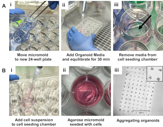
Figure 5: Schematic of ovarian somatic organoid generation. (A) Representative images of (i) transferring agarose micromolds to the culture plate with Graefe forceps, (ii) equilibration of agarose micromolds in Ovarian Organoid Media, and (iii) removal of media from cell seeding chambers. Arrow in Aiii indicates empty cell seeding chamber. (B) Representative images of (i,ii) seeding of cells into agarose micromolds and (iii) a transmitted light scan of an agarose micromold containing aggregating ovarian somatic cells. Scale bar (Biii): 2500 µm. Please click here to view a larger version of this figure.
5. Utilization of organoids for downstream applications
NOTE: Organoids can be utilized for a variety of downstream applications, including histology, molecular biology applications (e.g., RNA/protein analyses), conditioned media analysis, and co-culture.
- Histology
NOTE: Organoids can be processed for histological endpoints without removal from the agarose micromolds. In fact, organoids within agarose micromolds act similarly to tissue microarrays, thus allowing for high-throughput histologic assessment of several, consistently spaced organoids at one time.- Dissolve 1.5% (w/v) sterile agarose in PBS by boiling.
- Following the culture of ovarian somatic organoids, hold the plate at an angle and remove the media surrounding the agarose micromolds.
- Under a dissecting microscope, remove the media from the cell seeding chambers by gently hovering a pipette tip over the corner of each cell seeding chamber and carefully drawing up the media without disturbing the organoids or disrupting the micromold itself.
- Carefully pipette 100–125 µL of liquid 1.5% agarose cooled to approximately 50 °C into the corner of each cell seeding chamber to fill and seal the cell seeding chambers, taking care not to disturb the organoids (Figure 6Ai).
- Allow the agarose to solidify for 2–3 min at room temperature.
- Add 750 µL of Modified Davidson's or 4% paraformaldehyde to each well and fix the organoids within the sealed agarose micromolds overnight at 4 °C.
- After fixation, remove the fixative and wash the agarose micromolds with 70% ethanol.
NOTE: Micromolds can be stored in 70% ethanol at 4 °C until tissue processing (dehydration, clearing, and wax infiltration). During tissue processing, keep track of the orientation of the agarose micromolds. - Following tissue processing, embed the agarose micromolds in paraffin such that the micromolds are flat within the paraffin blocks and that the bottoms of the micromolds are the first to be sectioned through (Figure 6Aii).
- Section paraffin blocks at 5 µm and place the tissue sections on charged microscope slides.
NOTE: The presence of microwells can be visualized by eye during sectioning both in tissue sections and in paraffin blocks (Figure 6Aiii,iv). The presence of organoids within micromold sections can be verified by examining sectioned slides under a microscope (Figure 6Av). Rather than placing 2–3 consecutive sections on the same slide, placing 2–3 sections that are each 50 µm apart (i.e., every 10th section) onto a single slide increases the probability that most organoids will be represented in at least one section on each slide. Organoids are consistently spaced within tissue sections, increasing the ease of imaging following histological staining (Figure 6Avi). - Allow slides to dry in an oven at 37 °C overnight.
- Proceed with histological or immunohistochemical staining or store slides at room temperature19.
- Harvesting organoids for molecular applications
NOTE: Following culture, organoids can be removed from the agarose micromolds and processed for a variety of downstream molecular biology applications, including RNA or protein extraction to assess gene or protein expression.- Hold the plate containing the agarose micromolds at a slight angle and remove the media surrounding the agarose micromolds by pipetting.
- Add 750 µL of room temperature PBS to the wells containing the agarose micromolds.
- Use Graefe forceps to invert and agitate the agarose micromolds to release the organoids into the surrounding PBS. Confirm the release of the organoids from the agarose micromolds under a microscope.
- Remove the empty agarose micromolds using Graefe forceps and transfer the PBS containing the organoids into microcentrifuge tubes by pipetting.
- Pellet the organoids by centrifugation at 10,000 × g for 5 min at room temperature.
- Remove the supernatant by aspirating and proceed to RNA or protein extraction. Alternatively, flash-freeze the cell pellet and store at -80 °C19.
- Saving organoid conditioned media for analysis
NOTE: Conditioned media from organoids can be saved throughout culture for the analysis of organoid secreted factors, including hormones, cytokines, and growth factors. Representative results from analysis of cytokines in organoid conditioned media are shown in Figure 7A.- Hold the plate containing the agarose micromolds at a slight angle and remove the conditioned media surrounding the micromolds by pipetting. Do not remove media from the cell seeding chambers to avoid disturbing organoids.
- Transfer the conditioned media into microcentrifuge tubes and proceed to media analysis or flash-freeze and store at -80 °C19,22.
NOTE: Optionally, centrifuge the conditioned media at 10,000 × g for 5 min to pellet any dead cells or debris. Transfer the supernatant to new microcentrifuge tubes prior to media analysis or storage.
- Organoid co-culture
NOTE: Ovarian somatic organoids can be co-cultured with other cells and tissues by several methods. Representative results from organoid co-culture with ovarian cancer cells and follicles, by the methods described in steps 5.4.1 and 5.4.3, respectively, are shown in Figure 7B,C.- To generate organoids using a combination of primary mouse ovarian somatic cells and other cell types to investigate the effects of direct cell-cell interactions, follow the steps 5.4.1.1–5.4.1.4.
- Follow steps 4.1–4.9 to wash primary ovarian somatic cells following 2D culture overnight, harvest the cells from 2D culture using 0.05% Trypsin, pellet, count, and resuspend the cells in ovarian organoid media at a density of 2.5 × 10⁵ live cells per 75 µL of media.
- If the other cell type of interest is adherent, harvest the cells from a 2D culture. Count the cells, pellet them by centrifugation, and resuspend in ovarian organoid media at a density of 2.5 × 10⁵ live cells per 75 µL of media.
- Combine the suspension of primary ovarian somatic cells with the suspension of the other cell type of interest at a known ratio.
- Follow steps 4.10–4.13 to seed the resulting cell suspension into agarose micromolds and culture organoids.
- To transfer agarose micromolds containing formed organoids to 24-well plates containing other cells or organoids, follow steps 5.4.2.1–5.4.2.4.
NOTE: To assess the effects of organoid-derived secreted factors on other cell types and vice versa, formed organoids can remain within the agarose micromolds, and entire agarose micromolds can be transferred to a 24-well plate containing other cells or organoids. These co-culture methods can be utilized when ovarian organoid media can be used to culture both cell/tissue types.- Warm ovarian organoid media to 37 °C in a water bath.
- Hold the plate containing the agarose micromolds at an angle and remove the media surrounding the agarose micromolds by pipetting.
- Using Graefe forceps, carefully transfer the micromolds into 24-well plates containing other cells or organoids.
- Add 750 µL of fresh ovarian organoid media to surround the agarose micromolds.
- To remove formed organoids from the agarose micromolds and transfer them to new agarose micromolds, follow steps 5.4.3.1–5.4.3.13.
NOTE: If ovarian organoid media is not the desired media for co-culture experiments, the paradigm described in step 5.4.2 poses the challenge of significant media carryover, given that agarose micromolds are saturated with ovarian organoid media. Formed organoids (1-3 days following initial cell seeding) can be moved from their agarose micromolds and transferred into new agarose micromolds for additional culture or co-culture in order to prevent media carryover.- Warm the desired media for co-culture to 37 °C in a water bath.
NOTE: DMEM, αMEM, and F12-based media have successfully been used for co-culture experiments with ovarian somatic organoids. - Using Graefe forceps, carefully transfer the new agarose micromolds from PBS +1% PS into wells of a 24-well plate containing other cells or organoids for co-culture.
- Make sure that micromolds are fully submerged in media, add additional desired media for co-culture as necessary, and equilibrate the micromolds and media for at least 30 min in an incubator (humidified environment, 37 °C with 5% CO2).
- Hold the plate containing the agarose micromolds with formed organoids at a slight angle and remove the media surrounding the agarose micromolds by pipetting.
- Add the desired media for co-culture surrounding the agarose micromolds with the formed organoids.
- Use Graefe forceps to invert and agitate the agarose micromolds to release the organoids into the surrounding media. Confirm the release of organoids from the agarose micromolds under a microscope.
NOTE: If primary ovarian somatic cells have not aggregated adequately, organoids may not remain intact when removed from agarose micromolds. Organoid assembly may take 1–3 days, depending on the strain and age of the mice from which ovarian somatic cells were isolated. - Remove the empty agarose micromolds using Graefe forceps and transfer the media containing the organoids into microcentrifuge tubes by pipetting.
- Centrifuge the organoids at 300 × g for 5 min at room temperature.
- Under a dissecting microscope, remove the supernatant by pipetting, leaving 50–75 µL of media containing the organoids.
- Remove the equilibrated micromolds from the incubator and make sure that the micromolds are oriented such that the cell seeding chamber is facing up.
- Remove the media from the cell seeding chambers using Graefe forceps or by pipetting.
NOTE: Wells should not contain more than 500 µL of media so that the cell seeding chambers of micromolds can be emptied. - Transfer the organoids directly to the cell seeding chamber of each new agarose micromold by pipetting. Return the 24-well plate to the incubator.
- After 1–2 h, add an additional 250 µL of the desired media around the outside of agarose micromolds, being careful not to disturb the cell seeding chambers.
- Warm the desired media for co-culture to 37 °C in a water bath.
- To generate organoids using a combination of primary mouse ovarian somatic cells and other cell types to investigate the effects of direct cell-cell interactions, follow the steps 5.4.1.1–5.4.1.4.
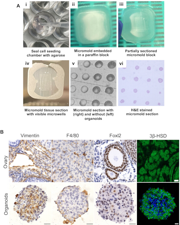
Figure 6: Key ovarian cell types are preserved in ovarian somatic organoids. (A) Representative images of (i) sealing a micromold with agarose at the end of culture, (ii) an uncut micromold, and (iii) a sectioned micromold embedded in a paraffin block. Microwells can be visualized in (iv) mounted sections, and the presence of organoids within sectioned micromolds can be (v) confirmed under a microscope prior to (vi) histological staining. (B) Representative images of mouse ovarian tissue sections and ovarian somatic organoid sections following immunohistochemical staining using antibodies against Vimentin, F4/80, Foxl2, and 3β-HSD. Scale bars = 20 µm. Abbreviations: H&E = hematoxylin and eosin. This figure has been adapted with permission from Dipali et al.19. Please click here to view a larger version of this figure.
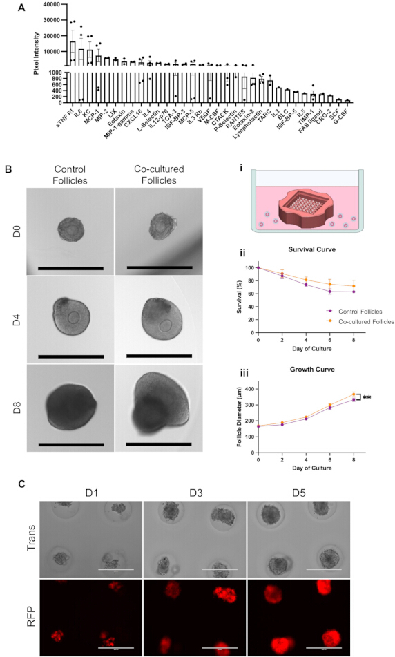
Figure 7: Co-culture of ovarian somatic organoids with follicles and ovarian cancer cells. (A) Organoid cytokine secretion was measured per agarose micromold at day 5 of culture and was normalized to conditioned media from agarose micromolds without organoids. N = 4 agarose micromolds. (B) Representative transmitted light images of ovarian follicles at days 0, 4, and 8 of culture with or without (control) organoids and (i) a schematic of the co-culture paradigm. Scale bars = 400 µm. Quantification of (ii) follicle survival and (iii) growth over 8 days of culture with or without (control) organoids. Data are shown as mean ± SD. Some error bars are too small to be visualized. N = 56 follicles per group. **P < 0.01. (C) Representative transmitted light (top) and fluorescent (bottom) images of primary ovarian somatic cells cultured at a 10:1 ratio with red fluorescent protein-tagged ovarian cancer cells (OVCAR8-RFP) in organoids at days 1, 3, and 5 of culture. Scale bars = 400 µm. Abbreviation: RFP = red fluorescent protein. Please click here to view a larger version of this figure.
6. Troubleshooting tips
- Problem 1: The number of isolated primary ovarian somatic cells is lower than anticipated.
NOTE: If ovarian tissue fragments are consistently smaller than 1 mm or not visible by eye following enzymatic digestion, the isolated cell number may be low because of decreased viability due to over-digestion. If ovarian tissue fragments are larger than 1 mm following enzymatic digestion, the isolated cell number may be low because of insufficient digestion of the ovarian tissue.- If ovarian tissue fragments are larger than 1 mm following enzymatic digestion, follow steps 6.1.1.1–6.1.1.2.
- Confirm that Collagenase IV and DNase I are within their recommended shelf-life and have not lost enzymatic activity due to improper storage or, in the case of DNase I, repeated freeze-thaw cycles.
- Increase the amount of repeated pipetting of the ovarian tissue, decrease the bore size of the p1000 tip if it was cut to make the opening wider, and/or increase the incubation time in EM.
- If ovarian tissue fragments are consistently much smaller than 1 mm following enzymatic digestion, decrease the amount of repeated pipetting of the ovarian tissue, increase the bore size of the p1000 tip by cutting to make the opening wider, and/or decrease the total incubation in EM.
- If ovarian tissue fragments are larger than 1 mm following enzymatic digestion, follow steps 6.1.1.1–6.1.1.2.
- Problem 2: Greater than ~25% of casted agarose micromolds have defects
- Confirm that the silicone cast is free of any residual agarose and debris. If needed, wash the silicone casts in PBS and re-sterilize by autoclaving. If needed, re-heat the 1% agarose used for casting, as agarose that is solidifying may not uniformly fill the silicone cast and/or result in bubbles.
- Once pipetted into the silicone cast, allow agarose to solidify completely, as premature removal of the agarose micromold from the cast can result in disrupted microwells.
- Confirm that silicone casts are not underfilled, as this can reduce the structural integrity of the agarose micromold, leading to damage when the silicone is flexed to remove the agarose micromold.
- Additionally, the manufacturer of the silicone casts recommends that they be autoclaved no more than 12 times. If the silicone cast has undergone excessive sterilization, replace it.
- Problem 3: Organoids fail to form in greater than 10% of microwells
- Confirm that the silicone casts are not overfilled when casting the agarose micromolds, as this can lead to irregular distribution of cells into the microwells, favoring the central microwells in the agarose micromold over the outer microwells.
- Avoid creating bubbles when dispensing the cell suspension into the cell seeding chamber of the agarose micromolds, as bubbles can disrupt the distribution of cells into microwells.
- If needed, start with greater than 75 µL cell suspension (at a density of 3.33 × 10⁶ live cells per 1 mL) per micromold to ensure that bubbles are not created due to insufficient volume in the pipette tip.
- Increase the interval between cell seeding and the addition of ovarian organoid media to the outside of the agarose micromold to ensure sufficient time for cells to settle into microwells.
Access restricted. Please log in or start a trial to view this content.
Results
Loading...
$$\rightleftharpoonup{xx}$$
$$\longleftharp{xx}$$,
$$\longrightharp{xx}$$,
To determine the cellular composition of ovarian somatic organoids, we performed immunohistochemistry of organoid tissue sections after 6 days of culture using antibodies against key ovarian cell types. Organoids contained vimentin-positive fibroblasts, F4/80-positive macrophages, as well as steroidogenic cell populations (Figure 6B)19. We utilized an antibody against Foxl2 to mark granulosa and granulosa-lutein cells, in addition to an antibody against 3β-HSD to mark...
Access restricted. Please log in or start a trial to view this content.
Discussion
Loading...
$$\rightleftharpoonup{xx}$$
$$\longleftharp{xx}$$,
$$\longrightharp{xx}$$,
This paper describes methods to isolate primary mouse ovarian somatic cells from a stroma-enriched fraction of the ovary, in a manner that maintains the heterogeneity of the ovarian somatic compartment. Primary mouse ovarian somatic cells can be cultured in a 2D monolayer or can be utilized to generate ovarian somatic organoids using scaffold-free, agarose micromolds. Agarose micromolds contain 96 microwells for organoid formation and fit in a single well of a 24-well plate, making this a high-throughput method to genera...
Access restricted. Please log in or start a trial to view this content.
Disclosures
Loading...
$$\rightleftharpoonup{xx}$$
$$\longleftharp{xx}$$,
$$\longrightharp{xx}$$,
The authors have no conflicts of interest to disclose.
Acknowledgements
Loading...
$$\rightleftharpoonup{xx}$$
$$\longleftharp{xx}$$,
$$\longrightharp{xx}$$,
This work was funded by the National Institute of Child Health and Human Development (R01HD093726 to F.E.D. and T32HD094699 to S.S.D. and E.J.Z.), the National Cancer Institute (F31CA257300 to S.S.D.), and startup funds from the Department of Obstetrics and Gynecology (to F.E.D.). We want to acknowledge Dr. Candace Tingen and the laboratory of Dr. Teresa Woodruff, as well as Dr. Jennifer Rowley, for their foundational studies and optimization of methods to isolate primary mouse ovarian somatic cells and characterization of these cells.
Access restricted. Please log in or start a trial to view this content.
Materials
| Name | Company | Catalog Number | Comments |
|---|---|---|---|
| 0.2 μm, Polyethersulfone syringe filter | Corning | 431229 | |
| 1.5 mL microcentrifuge tubes | MIDSCI | PR-MCT15 | |
| 100 mL glass beaker | Fisherbrand | FB100100 | |
| 100 mm Petri dishes | Corning | 351029 | |
| 15 mL conical tubes | ThermoFisher Scientific | 339651 | |
| 24-well plates (TC-treated) | Corning | 353047 | |
| 28-gauge insulin needles | Exel International | 26027 | |
| 35 mm Petri dishes | Corning | 351008 | |
| 3β-HSD antibody | Cosmo Bio | K0607 | 1:1000 |
| 4% Paraformaldehyde | Electron Microscopy Sciences | 1574100 | |
| 40 µm cell strainer | Corning | 431750 | |
| 45 mm watch glass | Electron Microscopy Sciences | 7054345 | |
| 50 mL concial tubes | ThermoFisher Scientific | 339652 | |
| 60 mL sterile syringe | Air-Tite | ML60 | |
| 60 mm TC-treated plates | Corning | 353037 | |
| 6-well plates (TC-treated) | Corning | 353046 | |
| Agarose | Hoefer | GR140-500 | Gel strength >1200 g/cm2; Gel Temperature 36 °C |
| Autoclave sterilization pouch | Fisherbrand | 01-812-55 | |
| Biotinylated Goat Anti-Mouse IgG | Vector Laboratories | BA-9200-1.5 | 1:200 |
| Biotinylated Goat Anti-Rabbit IgG | Vector Laboratories | PK-6101 | 1:200 |
| Biotinylated Goat Anti-Rat IgG | Vector Laboratories | BA-9400-1.5 | 1:100 |
| Bluing reagent | Fisherbrand | 23-245681 | |
| Centrifuge with swing bucket rotor for 50 mL conical tubes | ThermoFisher Scientific | Sorvall X1R Pro-MD | |
| Class II biosafety cabinet | The Baker Company | SG403A | |
| Cleaved Caspase-3 (CC3) antibody | Cell Signaling Technology | 9579T | 1:250 |
| CO2 incubator | ThermoFisher Scientific | Heracell VIOS 160i CO2 Incubator | |
| Collagenase, Type IV, powder | Gibco, Fisher Scientific | 17-104-019 | |
| C-Series Mouse Cytokine Antibody Array 3 Kit | RayBiotech | AAM-CYT-3-8 | |
| Cytoseal XYL | Epredia | 83124 | |
| Deoxyribonuclease I from bovine pancreas | Millipore Sigma | DN25-100MG | Dnase I |
| Dissecting microscissors | World Precision Instruments | WPI-14003 | |
| Dissecting scissors | World Precision Instruments | WPI-15922 | |
| Dissection microscope | Leica | MZ9.5, S9 series | |
| DPBS, no calcium, no magnesium | Gibco, Fisher Scientific | 14-190-250 | pH 7.0–7.3 |
| Eosin | Fisherbrand | 23-314631 | |
| F4/80 antibody | Bio-Rad | MCA497G | 1:50 |
| Fetal Bovine Serum, certified, heat inactivated, United States | Gibco, ThermoFisher Scientific | 10082147 | |
| Fine-tipped dissecting forceps | World Precision Instruments | WPI-14098 | |
| FOXL2 antibody | Abcam | ab246511 | 1:200 |
| Graefe Forceps | Fine Science Tools | 1105010 | |
| Hematoxylin | EK Industries | EKI 47971GL | |
| Hemocytometer | MedOne | DHCN015 | |
| Imaging System | ThermoFisher Scientific | EVOS FL Auto | |
| IntestiCult Organoid Growth Medium (Mouse) | STEMCELL Technologies | 06005 | Basal medium, Supplements 1 and 2 |
| Laminar flow hood | IVFtech | IVFtech sterile workstation | |
| Leibovitz's L-15 Medium | Gibco, Fisher Scientific | 11415114 | |
| Microscope slides | Mercedes Scientific | MER 7255/90/WH/CC | |
| MicroTissues 3D Petri Dish micro-mold spheroids | Millipore Sigma | Z764043-6EA | Silicone casts for agarose micromolds |
| Modified Davidson's Fixative | Electron Microscopy Sciences | 6413350 | |
| OVCAR8-RFP cells | Gift from Joanna Burdette PhD, University of Illinois Chicago | ||
| Penicillin-Streptomycin | Millipore Sigma | P4333-100ML | |
| Picrosirius Red staining solution | StatLab | STPSRPT | |
| RPMI 1640 Medium (ATCC modification) | Gibco, Fisher Scientific | A1049101 | |
| Tabletop centrifuge | Eppendorf | Centrifuge 5430 R | |
| Trypan blue | ThermoFisher Scientific | T10282 | |
| Trypsin-EDTA (0.05%), phenol red | Gibco, ThermoFisher Scientific | 25300054 | |
| Vimentin antibody | Cell Signaling Technology | 5741S | 1:100 |
| αMEM, GlutaMAX Supplement, no nucleosides | Gibco, Fisher Scientific | 32561102 |
References
Loading...
$$\rightleftharpoonup{xx}$$
$$\longleftharp{xx}$$,
$$\longrightharp{xx}$$,
- Thomas, F. H., Vanderhyden, B. C. Oocyte-granulosa cell interactions during mouse follicular development: regulation of kit ligand expression and its role in oocyte growth. Reprod Biol Endocrinol. 4 (1), 19(2006).
- Robker, R. L., Hennebold, J. D., Russell, D. L. Coordination of ovulation and oocyte maturation: a good egg at the right time. Endocrinology. 159 (9), 3209-3218 (2018).
- Casey, G. Sex hormones and health. Nurs N Z. 23 (1), 24-28 (2017).
- Kinnear, H. M., et al. The ovarian stroma as a new frontier. Reproduction. 160 (3), R25-R39 (2020).
- Simon, L. E., Kumar, T. R., Duncan, F. E. In vitro ovarian follicle growth: a comprehensive analysis of key protocol variables. Biol Reprod. 103 (3), 455-470 (2020).
- Spector, I., Derech-Haim, S., Boustanai, I., Safrai, M., Meirow, D. Anti-Müllerian hormone signaling in the ovary involves stromal fibroblasts: a study in humans and mice provides novel insights into the role of ovarian stroma. Hum Reprod. 39 (11), 2551-2564 (2024).
- Kedem, A., et al. Activated ovarian endothelial cells promote early follicular development and survival. J Ovarian Res. 10 (1), 64(2017).
- Converse, A., et al. Multinucleated giant cells are hallmarks of ovarian aging with unique immune and degradation-associated molecular signatures. PLoS Biol. 23 (6), e3003204(2025).
- Bafor, E. E., et al. Isolation of single cells from individual mouse ovaries for flow cytometry and functional analysis. STAR Protoc. 4 (4), 102710(2023).
- Shepherd, T. G., Thériault, B. L., Campbell, E. J., Nachtigal, M. W. Primary culture of ovarian surface epithelial cells and ascites-derived ovarian cancer cells from patients. Nat Protoc. 1 (6), 2643-2649 (2006).
- Beschta, S., et al. A rapid and robust method for the cryopreservation of human granulosa cells. Histochem Cell Biol. 156 (5), 509-517 (2021).
- Tingen, C. M., et al. A macrophage and theca cell-enriched stromal cell population influences growth and survival of immature murine follicles in vitro. Reproduction. 141 (6), 809-820 (2011).
- O'Mara, A. E., et al. Chronic mirabegron treatment increases human brown fat, HDL cholesterol, and insulin sensitivity. J Clin Invest. 130 (5), 2209-2219 (2020).
- Zink, N., Stock, A. -K., Vahid, A., Beste, C. On the neurophysiological mechanisms underlying the adaptability to varying cognitive control demands. Front Hum Neurosci. 12, 411(2018).
- Zhao, Z., et al. Organoids. Nat Rev Methods Primers. 2 (1), 94(2022).
- Yang, S., et al. Organoids: the current status and biomedical applications. MedComm. 4 (3), e274(2023).
- Alzamil, L., Nikolakopoulou, K., Turco, M. Y. Organoid systems to study the human female reproductive tract and pregnancy. Cell Death Differ. 28 (1), 35-51 (2021).
- Pierson Smela, M. D., et al. Directed differentiation of human iPSCs to functional ovarian granulosa-like cells via transcription factor overexpression. eLife. 12, e83291(2023).
- Dipali, S. S., et al. Self-organizing ovarian somatic organoids preserve cellular heterogeneity and reveal cellular contributions to ovarian aging. Aging Cell. 25 (1), e70333(2026).
- Di Nisio, V., et al. Silk-ovarioids: establishment and characterization of a human ovarian primary cell 3D-model system. Hum Reprod Open. 2025 (3), hoaf042(2025).
- Nikanfar, S., Leonel, E. C. R., Damdimopoulou, P., Flaws, J. A., Amorim, C. A. Effects of phthalate exposure on human ovarian extracellular matrix composition: insights from a 3D spheroid model. Environ Res. 279, 121797(2025).
- Briley, S. M., et al. Reproductive age-associated fibrosis in the stroma of the mammalian ovary. Reproduction. 152 (3), 245-260 (2016).
- Converse, A., Zaniker, E. J., Amargant, F., Duncan, F. E. Recapitulating folliculogenesis and oogenesis outside the body: encapsulated in vitro follicle growth. Biol Reprod. 108 (1), 5-22 (2023).
- Dean, M., Jin, V., Russo, A., Lantvit, D. D., Burdette, J. E. Exposure of the extracellular matrix and colonization of the ovary in metastasis of fallopian-tube-derived cancer. Carcinogenesis. 40 (1), 41-51 (2019).
- Charalambous, C., Webster, A., Schuh, M. Aneuploidy in mammalian oocytes and the impact of maternal ageing. Nat Rev Mol Cell Biol. 24 (1), 27-44 (2023).
- Rowley, J. E., et al. Low molecular weight hyaluronan induces an inflammatory response in ovarian stromal cells and impairs gamete development in vitro. Int J Mol Sci. 21 (3), 1036(2020).
- Amargant, F., Vieira, C., Pritchard, M. T., Duncan, F. E. Systemic low-dose anti-fibrotic treatment attenuates ovarian aging in the mouse. Geroscience. 47 (3), 3475-3495 (2024).
Access restricted. Please log in or start a trial to view this content.
Reprints and Permissions
Request permission to reuse the text or figures of this JoVE article
Request Permission