This protocol combines standardized seed surface sterilization with controlled germination to enable the reproducible isolation of tomato seed-associated microbiota during the seed-to-seedling transition. The approach enhances recovery of culturable microorganisms that are underrepresented in dry seeds and supports comparative culturomic studies during early seedling development.
Method Article
Isolation of Tomato Seed-Associated Microbiota through Controlled Germination and Comparative Culturing
Opens in a new tab
In This Article
Summary
Loading...
$$\rightleftharpoonup{xx}$$
$$\longleftharp{xx}$$,
$$\longrightharp{xx}$$,
Abstract
Loading...
$$\rightleftharpoonup{xx}$$
$$\longleftharp{xx}$$,
$$\longrightharp{xx}$$,
Surface sterilization is a critical yet frequently under-optimized step in studies aiming to isolate seed-associated microorganisms, as excessively harsh treatments can reduce or distort endogenous microbial populations. This protocol presents a reproducible approach for isolating culturable tomato seed-associated microbiota using surface-sterilization conditions based on previously validated methodologies, combined with controlled seed germination. Rather than defining universally optimal sterilization treatments, the approach applies standardized conditions that can be adjusted according to seed characteristics while minimizing bias and preserving endogenous microbial populations.
To enhance recovery of culturable microorganisms associated with the seed-to-seedling transition, the protocol incorporates a controlled germination step prior to microbial isolation. In contrast to conventional approaches based on the maceration of dry, non-germinated seeds, controlled germination promotes microbial activation during the seed-to-seedling transition under sterile conditions. Comparative colony-forming unit (CFU)-based analyses of equivalent biological replicates, each consisting of pooled material from 3–5 dry seeds or 3–5 germinated seedlings, revealed a marked shift in the culturable community following germination. Genera that were weakly represented in dry seeds became enriched after germination, whereas other taxa that were more abundant in dry seeds decreased in relative representation, supporting studies of early plant-microbe interactions during seedling establishment.
Across biological replicates, genera including Stutzerimonas, Stenotrophomonas, and Priestia were consistently enriched among isolates recovered from germinated seedlings, whereas Paenibacillus remained abundant across conditions but showed marked shifts in species-level composition. By integrating standardized sterilization conditions with controlled germination and comparative analysis of dry and germinated material, this protocol provides a robust and reproducible framework for the culturomic isolation of seed-associated microbiota with potential functional relevance during early seedling development and supports studies of early plant–microbe interactions during seedling establishment.
Introduction
Loading...
$$\rightleftharpoonup{xx}$$
$$\longleftharp{xx}$$,
$$\longrightharp{xx}$$,
Seeds harbor diverse microbial communities composed of bacteria and fungi associated with both internal tissues and external surfaces1,2,3. These microorganisms are increasingly recognized as integral components of plant biology because they can influence plant performance from the earliest developmental stages, including germination, seedling establishment, and responses to biotic and abiotic stress1,2,3. In ecological terms, seeds function not only as propagules for plant reproduction but also as reservoirs of microbial inoculum, making them a critical entry point for understanding how plant-associated microbiomes are initiated and maintained across the plant life cycle1,2,3.
Seed-associated microorganisms may constitute the first microbial inoculum encountered by the emerging plant and can therefore contribute to microbiome assembly during the seed-to-seedling transition1,3,4. This early inoculum may influence the microbial communities that subsequently colonize roots and other plant compartments, with consequences for plant establishment and later plant-microbe interactions1,4. In addition, part of the seed microbiota can be vertically transmitted through reproductive structures such as flowers, ovules, and pollen, allowing microbial populations to persist across plant generations4,5. This intergenerational continuity suggests that some seed-borne microorganisms may consistently contribute to plant fitness and adaptation.
Functionally, seed-associated microorganisms may affect host performance through several mechanisms, including the production of phytohormones, antimicrobial compounds, and other bioactive metabolites that modulate plant development or suppress competitors and pathogens5,6,7. Seed-transmitted microbial populations also appear to coexist through differential resource use and ecological partitioning, suggesting that the seed represents a selective habitat rather than a passive container of microbial diversity7. Collectively, these findings support the idea that seeds host microbial populations that may influence early plant development and represent candidates for downstream functional characterization2,3,5,7.
To investigate seed-associated microorganisms in a functionally informative and experimentally tractable way, it is necessary to recover viable microbial populations from seed material. In this context, microbial isolation provides access to living microorganisms that can subsequently be characterized experimentally and evaluated in downstream assays relevant to plant–microbe interactions. This is particularly important in seed systems, where seed-associated microorganisms may contribute to early plant establishment and represent candidates for functional screening and future applied use.
The recovery of seed-associated microorganisms is highly sensitive to sample preparation procedures, particularly surface sterilization, tissue processing, and the developmental stage of the sampled material. This sensitivity is especially relevant in seed systems because microbial biomass is often low, and small procedural differences can strongly affect both colony-forming unit counts and isolate composition. Among these variables, surface sterilization is one of the most critical and difficult to optimize. Sterilization is necessary to reduce epiphytic contamination prior to microbial isolation, yet excessive exposure to sterilants can reduce the viability or culturability of endogenous microorganisms, whereas insufficient sterilization may allow surface-associated taxa to dominate subsequent cultures8,9,10. Accordingly, the aim of sterilization is not to maximize chemical exposure, but to apply the mildest treatment that effectively suppresses surface-derived contamination while preserving recoverable internal microorganisms5,11.
Because sterilization efficiency depends on plant species, tissue structure, seed coat properties, and the associated microbial community, protocols should be selected from previously validated methodologies and then adapted cautiously to the system under study8,12,13. In low-biomass materials such as tomato seeds, this balance is particularly important: over-sterilization may sharply reduce microbial recovery, whereas under-sterilization increases the likelihood that fast-growing surface contaminants will dominate the isolates obtained. For this reason, sterilization efficiency should be verified routinely rather than assumed. Plating of the final rinse water provides a practical control for residual external contamination, but this control should always be interpreted together with microbial recovery from seed or seedling homogenates. A protocol that yields sterile rinse controls but little or no recoverable internal microbiota may be unsuitable when the objective is comparative microbial isolation rather than maximal decontamination6,8,10,14.
Meaningful comparisons, therefore, require strong procedural standardization. In low-biomass samples, inconsistencies in washing, transfer, homogenization, dilution, plating, or incubation can alter both total microbial recovery and the apparent representation of individual taxa. This is especially important when comparing dry and germinated material, where methodological variation can easily be mistaken for biological change.
A major limitation of many seed microbiology protocols is that they rely exclusively on dry, non-germinated seeds as starting material. Although this provides a useful baseline for the microbial fraction present before seed activation, it may underestimate microorganisms that are dormant, metabolically inactive, stressed, or initially present at low relative abundance2,15.
In the dry state, seeds represent a physiologically quiescent environment in which both host tissues and associated microorganisms may remain relatively inactive. Consequently, the microbial populations recovered directly from dry seeds may not fully represent those that become more readily recoverable once germination begins.
During germination, seeds undergo water uptake, metabolic reactivation, nutrient mobilization, and tissue reorganization, all of which alter the local environment available to associated microorganisms11,16. These changes can promote microbial growth, alter the relative representation of microbial populations, and facilitate the detection or recovery of taxa that were previously rare or poorly recovered in dry seeds. In addition, radicle emergence creates a new plant compartment that may support microbial proliferation and redistribution, making the seed-to-seedling transition a particularly informative stage for microbiological analysis1,17.
For this reason, parallel processing of dry seeds and germinated seedlings provides a more informative experimental design than analysis of dry seeds alone. Dry seeds establish a baseline for the culturable populations present before activation, whereas germinated material allows comparative assessment of taxa that become more readily recoverable during early development. This approach is particularly useful when the aim is not only to catalog recoverable microorganisms, but also to identify taxa associated with the seed-to-seedling transition and prioritize isolates for downstream characterization (Figure 1).
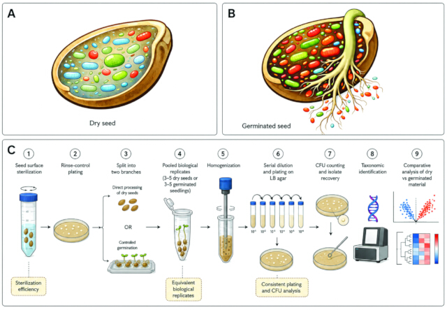
Figure 1: Conceptual and procedural overview of the comparative isolation workflow. (A) Conceptual representation of the dry tomato seed condition prior to germination. (B) Conceptual representation of the germinated tomato seed condition during the seed-to-seedling transition. (C) Schematic workflow of the protocol, including seed surface sterilization, rinse-control assessment, direct processing of dry seeds or controlled germination, homogenization of pooled biological replicates, serial dilution and plating, CFU-based quantification, isolate recovery, taxonomic identification, and comparative analysis of dry and germinated material. Critical control points highlighted in the workflow include sterilization efficiency, equivalent biological replicates, and consistent plating and CFU analysis. Panels A and B were prepared as AI-assisted conceptual illustrations for visual representation only. Please click here to view a larger version of this figure.
Because the protocol is intended to compare microbial recovery from dry and germinated material, strict methodological consistency is essential. All comparisons should be performed using the same seed lot, identical sterilization conditions, the same culture media, and equivalent dilution, plating, and incubation procedures. Without this level of standardization, apparent differences between dry and germinated samples may reflect technical variation rather than biologically meaningful enrichment. This is particularly important in isolation workflows, where the recovered fraction is shaped both by the biological sample and by the selective effects of the experimental procedure itself4,15.
Biological replication is likewise indispensable. Low-abundance taxa may vary among seed batches, and microbial recovery from low-biomass samples is inherently stochastic even under carefully controlled conditions. At least five independent biological replicates per condition are recommended for robust comparison. In this workflow, each biological replicate consists of pooled material from 3–5 dry seeds or 3–5 germinated seedlings processed as a single sample. In addition, all manipulations after sterilization should be performed under aseptic conditions to minimize secondary contamination and preserve interpretability of the resulting isolate set.
The duration of germination should also be selected carefully. It must be long enough to permit radicle emergence and early seedling development, but not so prolonged that later plant growth introduces additional ecological complexity unrelated to the immediate seed-to-seedling transition. For tomato, a germination period of up to 5 days under sterile conditions provides a practical window in which the seedling remains closely linked to the seed environment while early microbial activation and redistribution can already occur. Consistent timing across experiments is essential because differences in developmental stage may themselves influence microbial recovery patterns. This workflow is most appropriate for comparative recovery of culturable microbiota from low-biomass seed systems when the aim is to evaluate germination-associated shifts under standardized conditions. It is less suitable for direct estimation of total seed microbiome diversity or for experimental systems in which culture-independent approaches are required to capture the broader microbial community.
The objective of this protocol is to provide a reproducible framework for the isolation of culturable tomato seed-associated microbiota by integrating standardized surface sterilization with controlled seed germination. The method enables comparative analysis of dry seeds and germinated seedlings using equivalent biological replicates, each consisting of pooled material from 3–5 dry seeds or 3–5 germinated seedlings, to assess germination-dependent shifts in colony-forming unit (CFU) recovery and taxonomic composition. By minimizing sterilization bias and promoting microbial recovery during the seed-to-seedling transition, this approach facilitates the isolation of microorganisms with potential functional relevance during early seedling development and supports downstream studies of early plant–microbe interactions.
Access restricted. Please log in or start a trial to view this content.
Protocol
Loading...
$$\rightleftharpoonup{xx}$$
$$\longleftharp{xx}$$,
$$\longrightharp{xx}$$,
1. Preparation of reagents, media, and sterile materials
- Prepare a 70% (v:v) ethanol solution using sterile distilled water.
- Prepare a 20% (v:v) working solution from commercial bleach containing 5% available sodium hypochlorite by diluting 20 mL of bleach in 80 mL of sterile distilled water.
- Prepare sterile distilled water for washing steps.
- Prepare sterile 0.45% NaCl for homogenization and serial dilution, and use this solution consistently throughout the workflow.
- Prepare Luria Bertani (LB) agar culture medium (per liter: 10 g of tryptone, 5 g of yeast extract, 10 g of NaCl, and 15 g of agar) and pour plates in advance under sterile conditions.
- Sterilize all instruments that will contact seeds or seedlings after sterilization, including forceps, pestles, spreaders, scissors, and tube racks, by autoclaving or validated aseptic treatment.
- Arrange sterile Petri dishes, tubes, pipette tips, and culture plates inside a laminar flow hood before beginning seed handling.
NOTE: Because seed-associated microbial biomass may be low, it is important to minimize unnecessary handling time and to have all materials ready before starting sterilization. Delays between chemical treatments, washing, plating, or germination setup may introduce variability in recovery. Alternative sterile buffers may be used in other experimental systems, but a single buffer should be maintained throughout any given comparative experiment. Additional media such as Reasoner's 2A (R2A) or selective media may be included in extended culturomic workflows, but LB agar was used as the standard medium for the comparative protocol described here.
2. Selection and allocation of seeds
- Use seeds from a single tomato seed lot for each experiment whenever possible.
NOTE: In the representative workflow described here, tomato seeds from a single seed lot of cv. Money Maker were used throughout each experiment. - Visually inspect seeds and discard damaged, visibly broken, or malformed seeds.
- Divide the seeds into the experimental groups required for the study. At minimum, allocate seeds to: dry-seed isolation after sterilization, and germination followed by seedling isolation.
- If more than one sterilization condition is to be tested, assign seeds randomly across sterilization treatments to avoid treatment bias caused by seed size or condition.
NOTE: All downstream comparisons are stronger when based on the same seed batch processed in parallel. The workflow may be adapted to other tomato genotypes or varieties, but any comparison within an experiment should be performed using a single seed lot processed in parallel.
3. Surface sterilization of tomato seeds
- Transfer 3–5 seeds into each sterile 2 mL microcentrifuge tube inside a laminar flow hood.
- Add 1 mL of 70% (v:v) ethanol to each tube to fully immerse the seeds.
- Incubate the tubes for 5 min with gentle mixing by tube rotator at 25 rpm.
- Remove the ethanol completely using a sterile pipette.
- Immediately add 1 mL of the 20% (v:v) sodium hypochlorite working solution to each tube to fully immerse the seeds.
- Incubate the tubes for 10 min with gentle agitation by the tube rotator at 25 rpm.
- Remove the sodium hypochlorite solution completely using a sterile pipette.
- Wash the seeds three times with 1 mL of sterile distilled water per wash.
- For each wash, add sterile water, mix gently for 30 s, allow the seeds to settle for 30 s if needed, and remove the wash solution completely before adding fresh sterile water.
- Retain the final rinse water from each tube for sterilization efficiency controls.
- Transfer the sterilized seeds to sterile Petri dishes or sterile tubes, according to whether they will be processed immediately as dry seeds or used for controlled germination.
CAUTION: Handle sodium hypochlorite using appropriate personal protective equipment and perform all steps according to institutional chemical safety procedures.
NOTE: The ethanol-sodium hypochlorite sequence used here was selected as a standardized working condition for tomato seeds based on previously validated sterilization workflows. Use the same sterilization conditions for all samples included in a comparative experiment. If more than one sterilization regime is evaluated, process each treatment in parallel and assess sterilization efficiency independently for each condition. Minimize the time between the final wash and downstream processing to reduce variability in microbial recovery.
4. Verification of sterilization efficiency
- Prepare 10-fold serial dilutions of the final rinse water in sterile 0.45% NaCl by transferring 100 µL of rinse water into 900 µL of diluent and repeating the same step sequentially up to 10-4. Plate 100 µL of each dilution onto LB agar using the same plating method applied to dry-seed and germinated-seedling homogenates.
- Incubate the plates at 28 °C for 24–48 h.
- Inspect the plates for colony growth after incubation.
- Record the presence or absence of colonies for each dilution. If colonies are present, record the dilution level, approximate colony number, and visible colony morphology.
NOTE: Use the same plating volume and plating format throughout the experiment to allow direct comparison between rinse controls and biological samples. If no colonies are detected in the undiluted or lowest-dilution rinse-control plates, record the sample as having no detectable growth under the culture conditions used.
5. Processing of dry (non-germinated) seeds
- Transfer pooled sets of 3–5 sterilized dry seeds, corresponding to one biological replicate, into sterile 2 mL microcentrifuge tubes containing 1 mL of sterile 0.45% NaCl.
- Homogenize the seeds using a sterile disposable pestle until the seed tissues are visibly disrupted and no intact seed structures remain.
- Vortex the homogenate for 30 s to resuspend microbial cells uniformly.
- Prepare 10-fold serial dilutions of the homogenate in sterile 0.45% NaCl by transferring 100 µL of homogenate into 900 µL of diluent for the first dilution and repeating the same ratio sequentially up to 10-6.
- Plate 100 µL of the selected dilutions onto LB agar using spread plating.
- Incubate the plates at 28 °C for 24–72 h.
- Select the plates corresponding to the dilution range that yields isolated and countable colonies for downstream CFU estimation and isolate recovery.
- Record the total colony count, visible colony morphotypes, and any differences in growth pattern among replicates.
NOTE: Perform dry-seed processing in parallel with germinated-seedling samples using the same culture medium, dilution scheme, plating volume, and incubation conditions. LB agar was used as the standard medium for the comparative workflow described here. Additional media may be incorporated in extended isolation workflows when broader recovery of slow-growing taxa is required.
6. Axenic germination of sterilized seeds
- Place sterilized seeds on sterile germination support, such as Murashige and Skoog (MS) agar plates.
- Space seeds evenly to avoid contact between emerging seedlings.
- Seal plates with laboratory sealing film or equivalent material that reduces external contamination while allowing normal germination.
- Incubate the plates horizontally at 25 °C in the dark.
- Allow seeds to germinate under sterile conditions until early radicle establishment is clearly visible; in the representative experiment, this corresponded to 5 days of incubation.
- Inspect plates regularly for signs of contamination, abnormal germination, or excessive condensation.
NOTE: Germination conditions should be kept constant among replicates. Variation in germination duration or seedling stage may alter microbial recovery independently of the effect being studied. Other sterile germination supports, including Phytoagar plates or sterile moist filter paper, may be used in adapted workflows, provided that the support remains constant within each experiment. Alternative plate orientations may be used for incubation, depending on the germination container, but orientation should be kept constant within the experiment. (Pause point) Germinated seedlings may be held briefly at 4 °C before processing if necessary, but immediate processing is preferred whenever possible.
7. Isolation of microorganisms from germinated seedlings
- Collect germinated seedlings using sterile forceps inside a laminar flow hood.
- Transfer pooled material from 3–5 germinated seedlings, corresponding to one biological replicate, into sterile 2 mL microcentrifuge tubes.
- Add 1 mL of sterile 0.45% NaCl to each tube.
- Homogenize the pooled seedling material thoroughly using a sterile disposable pestle, ensuring that both the emerging root and the associated residual seed tissue are included, until the tissues are visibly disrupted.
- Vortex the homogenate for 30 s to release and resuspend microbial cells uniformly.
- Prepare 10-fold serial dilutions of the homogenate in sterile 0.45% NaCl by transferring 100 µL of homogenate into 900 µL of diluent for the first dilution and repeating the same ratio sequentially up to 10-4.
- Plate 100 µL of the selected dilutions onto LB agar using the sector, drop plating.
- Incubate the plates at 28 °C for 24–72 h. Record the total colony count, visible colony morphotypes, and differences relative to dry-seed samples.
NOTE: Define one biological replicate as pooled material from 3–5 germinated seedlings and process all replicates in parallel using the same dilution scheme, plating volume, culture medium, and incubation conditions. Include both residual seed tissue and the emerging root in the homogenate to maintain consistency across germinated-seedling samples.
8. Colony counting and isolate recovery
- After incubation, select the dilution plates that yield countable and well-separated colonies. Use plates containing 30–300 colonies for CFU estimation.
- Count the colonies on the selected plates and calculate CFU per biological replicate using the following formula:
CFU per replicate = (number of colonies × dilution factor)/plated volume (milliliter)- If more than one plate from the same dilution is used, calculate the mean colony count for that dilution before applying the formula.
- Record colony morphology using standardized descriptors, including size, shape, color, edge, elevation, and opacity.
- Select colonies for downstream isolation to minimize sampling bias. Select all distinguishable morphotypes when colony numbers are low, or randomly select multiple colonies across plates when colony numbers are high.
- Restreak the selected colonies onto fresh LB agar plates to obtain pure cultures.
- Repeat restreaking until each isolate shows a uniform colony morphology on the plate.
NOTE: Report clearly whether CFU values are expressed per biological replicate, per unit biomass, or using another normalization approach. If additional normalization approaches are applied, including biomass-based normalization, describe the calculation method explicitly in the Data analysis section.
9. Preservation of purified isolates
- Inoculate pure colonies into liquid LB culture medium and incubate until visible growth is obtained.
- Mix culture (1:1, v:v) with sterile 80% glycerol according to standard microbiological practice.
- Store cryostocks at -80 °C.
- Maintain a digital record for each isolate, including sample origin, replicate number, condition (dry seed or germinated seedling), sterilization treatment, date of isolation, plate medium, colony morphology, and downstream identification status.
NOTE: Accurate isolate metadata are essential if enrichment patterns are to be linked back to the seed stage, treatment, or biological replicate.
10. Molecular identification of isolates
- Extract genomic DNA from purified bacterial cultures using a bacterial DNA extraction kit according to the manufacturer's instructions.
- Amplify the bacterial 16S rRNA gene using either the V5-V8 hypervariable region or the near full-length 16S rRNA gene.
- For amplification of the V5–V8 region (~700 bp), use primers 799F (5′-AACMGGATTAGATACCCKG-3′) and 1392R (5′- ACGGGCGGTGTGTRC-3′).
- For amplification of the near full-length 16S rRNA gene (~1500 bp), use primers 27F (5′-AGAGTTTGATCCTGGCTCAG-3′) and 1492R (5′-GGTTACCTTGTTACGACTT-3′).
- Prepare each polymerase chain reaction (PCR) reaction in a final volume of 25 µL using PCR master mix, 0.4 µM of each primer, and 1 µL of template DNA.
- Run in a thermal cycler under the following conditions: initial denaturation at 95 °C for 2 min; 40 cycles of denaturation at 95 °C for 3 s, annealing at 45 °C for 30 s, and extension at 72 °C for 2 min; followed by a final extension at 72 °C for 7 min. Include a no-template control in each PCR run.
- Verify PCR products by electrophoresis on a 1% agarose gel and visualize the amplicons using a gel electrophoresis and imaging system.
- Purify the PCR products using a PCR clean-up kit according to the manufacturer's instructions and submit the purified amplicons for Sanger sequencing.
- Quality-check, trim, and assemble the resulting sequences where necessary.
- Compare each sequence against the National Center for Biotechnology Information (NCBI) nucleotide database using BLASTn (Megablast settings for highly similar sequences).
- Use the best-supported hit to assign taxonomy. Assign isolates to the genus level when the best hit shows adequate alignment coverage and at least 94.5% sequence identity.
- Use tentative species-level assignment only when the best hit shows at least 98.7% sequence identity, high query coverage, and no conflicting top matches among closely related species.
- If repeated amplification fails or sequence quality is insufficient, record the isolate as Unidentified. Because 16S rRNA similarity alone may not resolve closely related bacterial species, treat all species-level assignments as provisional and confirm them by phylogenetic or genome-based analyses when required.
- Report isolate identity at the highest supported taxonomic resolution. Use these identifications to validate morphology-based grouping and to prioritize representative isolates for downstream genomic and functional analyses.
NOTE: This molecular identification step is intended to support isolate classification and representative strain selection. It should not be used to infer community structure or quantitative abundance patterns across treatments.
11. Data analysis
- Perform the experiment with at least five biological replicates per condition.
NOTE: In this workflow, each biological replicate consists of pooled material from 3–5 dry seeds or 3–5 germinated seedlings processed as a single sample. - For each condition, quantify: (i) total CFU recovery, (ii) number of visible colony morphotypes, (iii) number of purified isolates, and (iv) taxonomic composition of the recovered culturable microbiota at the genus level and, where relevant, at the species level.
- Compare dry-seed and germinated-seedling samples to assess germination-dependent changes in total recovery and taxonomic composition.
- Calculate the relative representation of each taxon as the percentage of total recovered CFUs under each condition. Calculate fold changes using absolute CFU values.
- Analyze quantitative data using an appropriate statistical analysis software (e.g., GraphPad Prism [v11.0.2]). Report data as mean ± standard deviation (SD).
- For pairwise comparisons between dry-seed and germinated-seedling samples, use a two-tailed Student's t-test when data meet the assumptions of normality.
- When those assumptions are not met, use a Mann–Whitney U test. Consider differences statistically significant at p < 0.05.
- Report clearly whether each value shown corresponds to absolute CFUs, relative CFU-derived percentages, or fold changes calculated from absolute CFU values.
NOTE: Use the same calculation criteria throughout the study to allow direct comparison between dry-seed and germinated-seedling samples. When presenting taxonomic composition, indicate explicitly whether values are shown at the genus level or the species level.
12. Critical quality-control points and troubleshooting
- A sterile final rinse does not by itself demonstrate that the selected sterilization regime is optimal for the experimental objective. Limit external contamination while preserving recovery of culturable microorganisms from the internal or tightly seed-associated fraction. Interpret rinse-control results together with microbial recovery from dry-seed and germinated-seedling homogenates.
- Low recovery from both dry and germinated samples may indicate over-sterilization, excessively harsh homogenization, inappropriate culture conditions, or delayed sample processing. In these cases, verify that sterilization times were applied consistently, confirm that tissue disruption was sufficient but not excessive, and ensure that dilution, plating, and incubation were performed immediately and under the same conditions across samples.
- High contamination in rinse controls, or rapid overgrowth by a small number of colony types, may indicate insufficient sterilization or contamination introduced during post-sterilization handling. In these cases, review sterilization timing, washing steps, aseptic handling, and the organization of materials inside the laminar flow hood before repeating the experiment.
- Interpret differences between dry and germinated samples biologically only when germination stage, biological replicate structure, culture medium, dilution scheme, plating volume, and incubation conditions were standardized across the experiment.
- When working with low-biomass seed material, the apparent absence of particular taxa in one condition may also reflect undersampling rather than true absence. For this reason, perform consistent replicate processing and isolate-level identification to robustly interpret germination-associated shifts.
13. Saving samples for other approaches
- After verification of sterilization efficiency and before plating, transfer an additional 200 µL aliquot of each dry-seed or germinated-seedling homogenate into a sterile 1.5 mL microcentrifuge tube.
- Label each tube with the sample type, biological replicate number, treatment condition, and date of collection.
- Freeze the aliquots immediately and store them at -80 °C until use in downstream analyses.
- Use the stored homogenate material for complementary approaches such as DNA-based analyses, microscopy-based assays, or confirmation experiments requiring the same starting biological replicate.
NOTE: Store the aliquots before serial dilution and plating to preserve the original homogenate composition. Avoid repeated freeze–thaw cycles by preparing separate aliquots when multiple downstream analyses are planned.
Access restricted. Please log in or start a trial to view this content.
Results
Loading...
$$\rightleftharpoonup{xx}$$
$$\longleftharp{xx}$$,
$$\longrightharp{xx}$$,
Performance of surface sterilization and control of external contamination
Surface sterilization using standardized conditions based on previously validated methodologies resulted in effective reduction of external microbial contamination while preserving the recovery of culturable microorganisms from seed-derived material. Plating of the final rinse water showed no detectable colony growth, indicating effective removal of surface-associated microorganisms under the conditions used.
Access restricted. Please log in or start a trial to view this content.
Discussion
Loading...
$$\rightleftharpoonup{xx}$$
$$\longleftharp{xx}$$,
$$\longrightharp{xx}$$,
The results presented here demonstrate that the integration of standardized surface sterilization with controlled seed germination provides a robust framework for the culturomic isolation of seed-associated microbiota. A key outcome of this protocol is the ability to balance effective removal of external contaminants with preservation of endogenous microbial populations. This balance is critical in culture-based studies, where overly stringent sterilization can reduce microbial recovery, while insufficient sterilization ...
Access restricted. Please log in or start a trial to view this content.
Disclosures
Loading...
$$\rightleftharpoonup{xx}$$
$$\longleftharp{xx}$$,
$$\longrightharp{xx}$$,
The authors declare that they have no competing financial interests or conflicts of interest related to this work. AI-assisted conceptual illustrations are used in this manuscript.
Acknowledgements
Loading...
$$\rightleftharpoonup{xx}$$
$$\longleftharp{xx}$$,
$$\longrightharp{xx}$$,
The authors thank ITQB NOVA (NOVA University of Lisbon, Oeiras, Portugal) and GREEN-IT Research Unit for access to greenhouse facilities and supporting infrastructure. The authors also thank Semillas Fitó for kindly providing the tomato seeds used in this study, with special thanks to Dr. Narváez (Seed Technologist, Semillas Fitó). This work was supported by FCT - Fundação para a Ciência e a Tecnologia, I.P., through Green-it Bioresources for Sustainability R&D Unit (UID/04551/2025, DOI: 10.54499/UID/04551/2025; UID/PRR/04551/2025, DOI: 10.54499/UID/PRR/04551/2025) and LS4FUTURE Associated Laboratory (LA/P/0087/2020, DOI: 10.54499/LA/P/0087/2020).
Access restricted. Please log in or start a trial to view this content.
Materials
| Name | Company | Catalog Number | Comments |
|---|---|---|---|
| Agar | Sigma-Aldrich | 01916-500G | Component of LB agar medium; 15 g/L. |
| Autoclave | Lab1st | SV120H | Used to sterilize instruments/materials or validate aseptic treatment. |
| Bacteria DNA extraction kit | Promocell | PK-MB708-206-100 | DNA extraction |
| Colony counting/recording setup | OCS | B079Y5W57N | Needed to record CFUs, morphotypes, and plate differences; manuscript does not specify counter/model. |
| Cryostorage vials | Sigma-Aldrich | Z359033 | Implied for storage of glycerol stocks at -80 °C; not named explicitly but needed for preservation step. |
| Digital isolate record system | Not specified in manuscript | Not applicable | Used to record isolate metadata: origin, replicate, condition, sterilization treatment, isolation date, plate medium, morphology, identification status. |
| Ethanol | VWR | 83813440 | Used to prepare 70% v/v ethanol solution for seed surface sterilization. |
| Filter paper | Fisher Scientific | 11718772 | Optional alternative germination support. |
| Forceps/Tweezers | Sigma-Aldrich | 930229 | Used to transfer seeds and collect germinated seedlings. |
| Genomic DNA extraction kit | PromoCell | PK-MB708-206-100 | Used to extract genomic DNA from purified bacterial cultures; exact kit/method not specified. |
| Glycerol, sterile 80% | VWR | 24388364 | Mixed 1:1 v/v with liquid culture for cryostock preparation. |
| Laminar flow hood Bio48 | Faster | LAB-CAP-22 | Required for aseptic seed handling, sterilization transfers, and preparation of sterile materials. |
| LB agar medium | VWR | 846495000 | Standard medium used for comparative culturing; per liter: 10 g of tryptone, 5 g of yeast extract, 10 g of NaCl, 15 g of agar. |
| Microcentrifuge tubes | Ratiolab | RATI5615000 | Used for seed sterilization and sample processing. |
| MS medium | Sigma | M0404-10L | Mentioned as sterile germination support for axenic germination. |
| NZYTaq II 2× Master mix | Nzytech | MB35803 | PCR mix for amplification |
| Parafilm | Parafilm/Merck | HS234526A | Used to seal germination plates while reducing contamination. |
| Petri dishes | Labbox | PDIP-E9N-500 | Used for germination setup, plating, and handling sterilized seeds. |
| Phytoagar | Duchefa Biochemie | 9002-18-0 | Mentioned as an optional alternative sterile germination support. |
| Pipettes set and tips | Merck | EP3123000918-1EA | Used for removing sterilization solutions, washing, dilution, and plating. |
| R2A agar medium | HI-MEDIA | M1743-500G | Optional additional medium for extended culturomic workflows. |
| Sodium chloride, NaCl | Fisher Chemical | 10040460 | Used to prepare sterile 0.45% NaCl for homogenization and serial dilutions; also part of LB medium. |
| Sodium hypochlorite | Carlo Erba | 370323 | Used to prepare 20% v/v sodium hypochlorite solution for seed surface sterilization. Corrosive; requires PPE. |
| Sterile disposable pestles | Eppendorf | EP0030120973 | Used for homogenization of dry seeds and germinated seedlings. |
| Vortex mixer | Fisher Scientific | 15901137 | Used to resuspend microbial cells from homogenates. |
Reprints and Permissions
Request permission to reuse the text or figures of this JoVE article
Request Permission